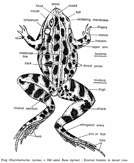
Dissection of Frog : External Features
For Dissection of Frog Procedure :- Note and examine the following in the living frog
- General body colour (adaptive colouration).
- Absence of neck and tail (helps in swimming).
- Hind limbs longer than forelimbs, which help in leaping and hopping.
- Up and down movements of the floor of the buccal cavity, showing respiratory movements.
- Distinguish between male and female frogs by copulatory pads in the hand of male.
For detailed external morphology, take a frog freshly killed with chloroform vapour. Draw a lateral or dorsal view of the frog. Examine and note the following
- Size :- Hoplobatrachus tigrinus (Rana tigrina) measures 12 to 20 em in length.
- Shape :- Roughly it is on triangular plane and adapted for swimming.
- Texture :- Skin is smooth, moist and slippery.
- Colouration :- The dorsal skin is green or olive coloured with dark patches, while ventral is whitish.
- Division :- Body is divided into two regions – head and trunk.
- Head :- Note the following structures in head which is conical and having blunt snout:
- External nares :- These are two small openings lying near the anterior tip of snout.
- Eyes. They are the most distinct parts of the head. Each eye is large, bright, protuberant, having golden iris and black rounded pupil. Each eye has an upper large and stationary eyelid and a lower small mobile eyelid.
- Nictitating membrane :- The upper part of lower eyelid forms a transparent nictitating membrane.
- Tympanum or tympanic membrane :- It is in the form of a circular area found posterior to each eye. It forms external boundary of the middle ear. The external ear is absent in frog. Vocal sacs. Found in males only under the head.
- Trunk :- It forms major portion of the body. Note the characteristic raised hump. Trunk contains anterior forelimbs and posterior hind limbs.
- Forelimbs :- Each comparises of upper arm, forearm and hand containing wrist, palm and four digits provided with copulatory and articular pads.
- Hind limbs :- These are folded at rest and each comparises of thigh, shank and foot. Foot is made up of ankle, sole and five digits.
- Head :- Note the following structures in head which is conical and having blunt snout:
Dissection of Frog : Bucco-pharyngeal Cavity
Procedure. Open the mouth of the frog as much as you can and note the various structures in the buccopharyngeal cavity.
- Mouth leads into buccal cavity, which is not sharply divided from pharyngeal cavity behind and hence they form common bucco-pharyugeal cavity formed by upper and lower jaws.
- Maxillary teeth are found in single row in the upper jaw.
- Vomerine teeth are found on the roof of upper jaw as two patch-like structures.
- Internal nares are found anteriorly in the mouth cavity as two small openings.
- Eye-balls bulge in from the roof of the upper jaw as two hemispherical structures.
- Openings of eustachian tubes are in the form of two circular openings found at the rear of buccal cavity.
- Tongue is long and bifid, attached anteriorly and free posteriorly.
- Gullet is found between two jaws and another opening called glottis which is found below the gullet.
- Vocal sacs are found in males having two lateral openings into the mouth cavity.
Dissection of Frog : Muscular System
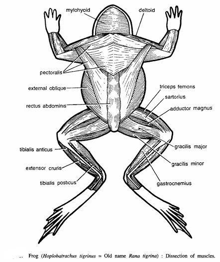
Procedure :- Remove the skin of the dorsal surface and note the following muscles
- Muscles on left side. Deltoid, triceps femoris, sartorius, adductor magnus, gracialis major, gracialis minor and gastronemius.
- Musscles on right side. Mylohyoid, pectoralis, external oblique, rectus abdominis, tibialis anticus extensor crusis and tibialis posticus.
Dissection of Frog : General Anatomy
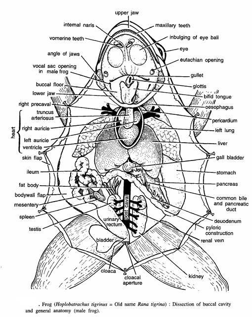
Procedure.
- Kill the frog with Chloroform vapours and lay it on its back in dissecting dish. Wash the frog, pin the limbs and press the pins with the base of forceps. Add water in the dish so that frog is completely submerged. Lift the abdominal skin with the forceps and make a mid-longitudinal incision from cloacal region up to the xiphoid cartilage and the pectoral girdle.
- Cut the skin of thighs to expose the muscles. Students generally cut or puncture the anterior abdominal vein, for which make two incisions on the sides of the abdominal skin. Ligate the anterior abdominal vein near the thighs and at its entrance into the liver and then cut between two ties. Examine and draw general viscera.
- Note that all the organs are connected by thin membranes called mesenteries. Study musculature, lungs, heart, liver, gall bladder, stomach, intestine, spleen, kidneys, fat-bodies, testes in male, ovaries and oviducts in female and urinary bladder.
Digestive system
- The inlet and outlet of the alimentary canal are mouth and cloaca. The mouth opening leads into the bucco-pharyngeal cavity which is followed by a short cylindrical oesophagus. The oesophagus opens into the stomach, which is muscular and curved lying on the left side of the abdominal cavity. It has cardiac and pyloric ends.
- The stomach opens into the intestine which comprises of small intestine and large intestine. The small intestine is called ileum and is thrown into several loops bound by mesenteries. The large intestine is short and straight and consists of the rectum which extends backwards and opens into the cloaca. The urinary bladder also opens into the cloaca.
- The accessory glands consist of liver having two unequal lobes. The two lobes are connected together by a median lobe or isthmus. The gall bladder lies between the two lobes. The hepatic ducts from the liver and cystic ducts from the gall bladder unite to form the common bile duct, which pierces the pancreas and accompanies the pancreatic duct, to open with it into the duodenum by a short common hepato-pancreatic duct near the pyloric end of the stomach.
Dissection of Frog : Circulatory System
Circulatory system of frog comprises of three units (i) a pumping organ or heart, (ii) the distributing vessels or arteries, and (iii) the collecting vessels or veins.
Heart of frog Procedure
For Dissection of Frog Remove pericardium to study the heart. The heart is S-shaped, triangular, conical and highly muscularised organ with broad anterior and narrow posterior regions. It is three-chambered, containing two auricles above and a conical ventricle below. The dorsal side of the heart is characterized by pulmonary veins, post-caval vessel and a reduced thin-walled sinus venosus. The ventral side contains a tubular truncus arteriosus.
Venous system of frog

Procedure
- Take preferably a freshly-chloroformed or killed frog, Pin it in the dissecting dish and open it with ventral surface upwards. The veins, in the dissecting or operating position, lie above arteries.
- Don’t pull unduly skin covering the thorax to the outside so as not to damage the musculo-cutaneous veins. Separate the anterior abdominal vein, tie it at both the ends and cut it in between. Separate alimentary canal from neighbouring organs by cutting mesenteries.
For renal portal system.
Dissection of Frog in the usual way. Separate the anterior abdominal vein and keep it intact. It is formed by the union of two pelvic veins lying in the anterior part of the pelvis. Try to separate the two pelvic veins from the thigh muscles by gently pulling the anterior abdominal vein forwards. Remove the vastus internus muscle lying on the outer side of the thigh to expose femoral vein. By scalpel cut through the pubic symphysis between the two thighs in order to expose the cloaca and two sciatic veins. De-oxygenated blood from all parts of the body is collected by the following :
- Pulmonary veins :- These collect oxygenated blood from lungs and open into left auricle.
- Caval veins :- The blood from rest of the viscera is poured by caval veins into the sinus venosus which, in turn, opens into the right auricle by a sinu-auricular aperture. The 3 caval veins are :
- Pre-cavals :- Two pre-cavals collect blood from anterior part of the body. Each pre-caval comprises of the following branches :
- External jugular. It divides into two veins. The lingual vein collects blood from tongue and floor of the mouth. The mandibular vein collects blood from lower jaw.
- Innominate. It also divides into two veins. The internal jugular collects blood from brain and orbit, while sub-scapular collects blood from shoulder and back arm.
- Sub-clavian. It has two branches. The branchial collects blood from the arm. The musculo cutaneous collects blood from muscles of abdomen and mucous membrane.
- Post-caval vein :- It receives blood from legs, kidneys, gonads and liver. It comprises of the following vessels :
- Femoral vein :- It collects blood from outer side of the leg.
- Sciatic vein :- It collects blood from inner side of the leg.
- Renal portal system :- Femoral vein divides into a renal portal vein and a pelvic vein. The sciatic vein joins with the renal portal vein, which runs on the outer side of kidney and enters kidney to break into several capillaries. The capillaries on emergence from kidney form 4 or 5 renal veins. The renal veins of two sides unite with post-caval vein.
- The double system of blood filteration, one in leg and another in kidneys, is called renal portal system. The renal portal vein also receives the dorso-lumbar vein, which collects blood from the back muscles. The two pelvic veins unite to form an anterior abdominal vein, which opens into the left lobe of the liver.
- Gonadial veins :- Each vein collects blood from a gonad and receives a small branch from the kidney. The gonadial veins join with the post-caval vein.
- Hepatic veins :- A pair of these, from the liver, joins with the post-caval vein.
- Hepatic portal system :- This system comprises of veins which collect blood from the alimentary canal and communicate with the large hepatic portal vein. A gastric vein collects blood from stomach, an intestinal vein from intestine, a duodenal vein from duodenum, a pancreatic vein from pancreas, and a splenic vein from spleen.
Arterial system of frog

The arteries distribute the oxygenated blood to various parts of the body.
Procedure
Dissection of Frog the freshly-killed frog as usual. Separate and tie both the ends of the anterior abdominal vein. Shift the alimentary canal to left hand side and pin it on the dissecting dish. Keep all the mesenteries intact. Tie the anterior vena cava. Lift the heart vertically, the anterior vena cava is seen on the dorsal side of the heart crossing over the three aortic arches of the same side.
Insert a fine thread on the anterior vena cava and after encircling it tie with the thread. Cut this vein a little ahead of the tied portion. Cut the pubic symphysis between the two thighs in order to expose the iliac arteries. Follow the branches of the truncus arteriosus. Truncus arteriosus divides into branches and each branch on each side gives rise to three main vessels
- Pulmo-cutaneous arch. It divides into two arteries :
- Pulmonary artery. It supplies to lung in which de-oxygenated blood is carried.
- Cutaneous artery. It supplies to skin and buccal cavity.
- Carotid arch. It divides into two arteries :
- External carotid. It supplies to tongue and hyoid.
- Internal carotid. It has a swollen carotid labyrinth and supplies to orbit and brain.
- Systemic arch. The two systemic arches encircle around the heart and join together to form the dorsal aorta. Each systematic arch gives the following branches :
- Oesophageal artery. It supplies to oesophagus.
- Occipito-vertebral artery. It supplies to head, backbone and spinal cord.
- Dorsal aorta. The dorsal aorta proceeds backwards and gives the following vessels
- Coeliaco-mesenteric artery. It divides into two. The coeliac artery supplies to stomach by gastric artery and to liver by hepatic artery. The anterior mesenteric vessel has 4 branches a duodenal supplying to duodenum, a splenic supplying to spleen, an intestinal, and intestine, and a posterior mesenteric supplying to large intestine.
- Gonadial artery. It supplies to gonad.
- Renal arteries. They supply to kidneys.
- Pelvic artery. It supplies to urinary bladder.
- Iliac arteries. Each iliac divides into two-a femoral artery supplying to outer side of the leg, and a sciatic artery supplying to inner side of the leg.
Dissection of Frog : Urinogenital System

Procedure
Dissection of Frog as usual. Separate the alimentary canal from oesophagus up to the rectum by cutting mesenteries and also cut pubic symphysis to expose the cloaca.
Male frog The urinary and genital organs function together. Urinary system includes :
- Kidney :- It is elongated and dark-red coloured with notched edges.
- Ureter :- It arises from outer side of kidney and opens into the cloaca. Upper part of seminal vesicle is called as ureter.
- Seminal vesicle :- It is formed by the dilation of ureter below kidney. Seminal continues backwards as urinogenital duct and opens into rectum and then into cloaca which opens to outside as cloacal aperture.
- Adrenal gland :- Ventral surface of each kidney has this yellow coloured, elongated gland.
- Urinary bladder :- It is a bilobed and thin-walled sac, opening into the floor of the cloaca. Genital system comprises of :
- A pair of testes :- Attached to anterior end of kidney by mesorchium.
- Fat bodies :- Finger-like bodies attached to each testis.
- Vasa efferentia :- About a dozen sperm ductules arise from each testis and communicate with collecting tubule of kidney, which opens into the ureter. Other structures seen are fat body, post-cava( veins and renal veins.
Female frog. The urinary and genital systems function independently and without any communication with each other. The urinary system is like that of male frog except that there is no dilation of ureter in the form of seminal vesicle. The genital system is on bilateral pattern. On each side it comprises of :
- Ovary :- Each ovary is lobulated and attached to the kidney by mesovarium. It contains countless ova.
- Ostium :- It is funnel-like opening of oviduct into coelom. The ostium leads into a swollen structure called as infundibulum which leads into oviduct.
- Oviduct. Oviduct is coiled tube and it opens into dilated uterus.
- Uterus. It is formed by posterior dilated oviduct. Uterus opens into the rectum. Rectum opens outside through cloacal aperture. Other structures seen are lung, urinary bladder and fat bodies.
Dissection of Frog : Cranial Nerves
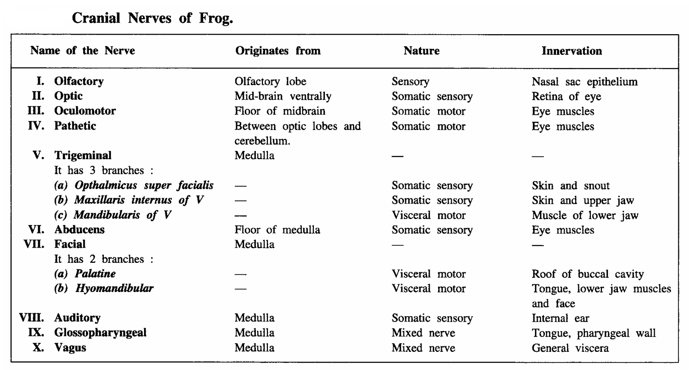
Procedure. Take anaesthesized frog. On one side remove the flap of skin around cranium upto lower jaw. From cranium 10 pairs of cranial nerves originate and innervate various organs as illustrated in the following chart. Trace the origin and innervation of the cranial nerves around optic lobe.
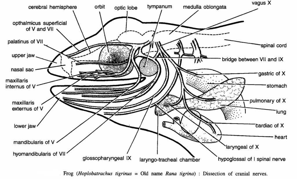
Image Refernces :- Practical zoology Vertebrate , Dissection of Frog