
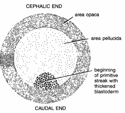
Chick Embryo – Whole Mount of 4 Hours of Incubation
Comments
- Four hours of incubation of the egg shows differentiation of the blastodisk into area pellucida and area opaca. Egg has cephalic end and caudal end.
- One quadrant of area pellucida becomes thickened, which marks the future caudal end of embryo.
- After 7 to 8 hours, the thickening becomes more elongated and represents start of primitive streak.
Identification : Since embryo shows beginning of primitive streak, hence it is chick embryo whole mount 4 hours of incubation.
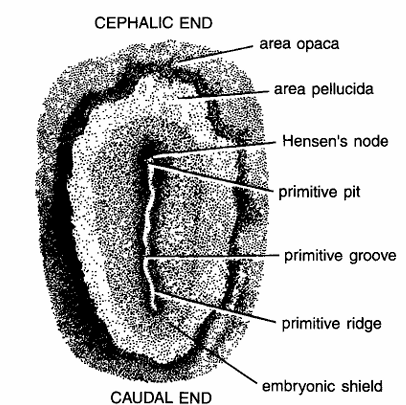
Chick Embryo – Whole Mount of 16 Hours of Incubation
Comments
- By 16 hours of incubation the primitive streak becomes so distinct that embryos are characterized as being in primitive streak stage
- In fixed and stained slide, w.m. primitive streak is composed of central furrow, called as primitive groove lined by thickened primitive ridges and ending in primitive pit.
- At the cephalic end of the primitive streak, closely-packed cells form thickened area, called as Hensen’s node.
- Part of area pellucida adjacent to the primitive streak shows increased thickness and forms embryonic area or embryonic shield. Area pellucida assumes elliptical shape.
- Elongated primitive streak represents long axis of future embryonic body.
- Caudal and cephalic ends are demacrated by Hensen’s node and end of primitive streak respectively.
Identification: Since it has primitive streak, hence it is whole mount of chick embryo after 16 hours of incubation.
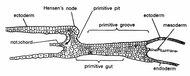
Chick Embryo : L.S. of 17 Hours of Incubation
Comments
- L.S. through 17 hours embryo represents the stage shortly after primitive streak formation and it also marks the beginning of morphogenetic movement of cells to form notochord.
- The section shows ectoderm, Hensen’s node, primitive pit, primitive groove, notochord and primitive gut and endoderm.
- The mesoderm extends on either side between ectoderm.
Identification : Since the section shows primitive pit and above features, hence it is L. S. of chick embryo of 17 hours of incubation.
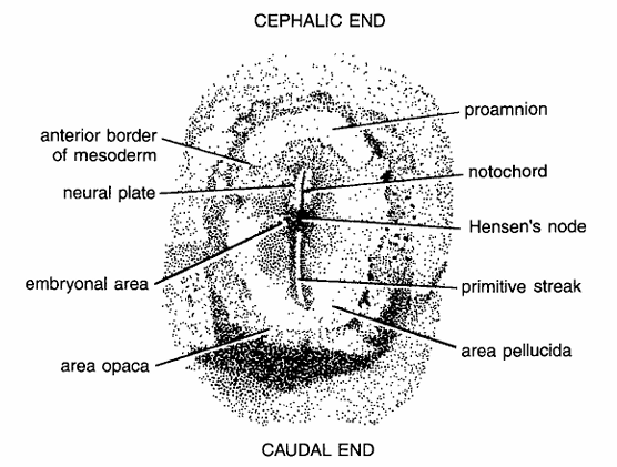
Chick Embryo : Whole Mount of 18 Hours of Incubation
Comments
- After 18 hours of incubation the notochord has become markedly elongated, forming a prominent structure
- Notochord extends towards cephalic region in the mid-line from Hensen’s node.
- 18 hours incubated embryo is spoken of as being in the head process stage.
- At the tip of the notochord neural plate develops.
- Caudal and cephalic ends are seen. In front of notochord and neural plate there is a space called as pro-amnion.
- Embryonic area, anterior border of mesoderm area pellucida and area opaca become more prominent.
- Primitive streak gradually decreases in size.
Identification : Since it has proamnion hence it is whole mount of chick embryo after 18 hours of incubation.
Chick Embryo : L.S. of 18 Hours of Incubation
Comments
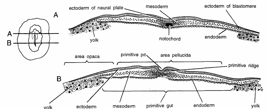
- Incubated embryo shows advanced inner structure.
- Ectoderm is shown by striated lines, the cells of mesoderm are represented by heavy angular dots and endoderm is represented by stippling backed by a single line.
- Section A shows yolk, ectoderm of neural plate, notochord, mesoderm, ectoderm and endoderm of blastoderm. Section B shows yolk, endoderm, primitive pit, primitive ridge, mesoderm and primitive gut, area pellucida and area opaca.
Identification : Since the sections shows neural plate (A) and primitive pit (B) hence they are sections of chick embryo L.S. after 18 hours of incubation.
Chick Embryo : Whole Mount of 24 Hours of Incubation

Comments
- At 24 hours of incubation the folding of the neural plate is much more clearly marked. In stained and transparent preparation of entire embryo neural folds appear as a pair of dark bands forming neural groove
- Neural folds at cephalic end are more prominent than at caudal end.
- Foregut is also established at this stage and the gut caudal to foregut called as midgut and the opening from midgut into foregut, namely anterior intestinal portal, also appears. In the middle, four pairs of somites are seen.
- The Hensen’s node is pushed caudally and primitive streak is further reduced.
- Other structures seen are area opaca vitellina, ectoderm of head, area pellucida, mesoderm, blood island, area vasculosa, notochord, mesenchyme, proamnion, Hensen’s node, sub-cephalic pocket and unsegmented mesoderm.
Identification : Since it has neural fold and above features hence it is whole mount of chick embryo of 24 hours of incubation.
Chick Embryo: T.S. Passing Through 24 Hours of Incubation
Comments
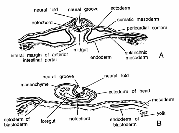
- T.S. passing through head
- T.S. passing through head region shows neural plate folded to form a complete neural tube.
- Beneath neural fold is notochord.
- Other structures seen in section are mesenchyme, foregut, ectoderm of head, mesoderm and endoderm of blastoderm and ectoderm of blastoderm.
- T.S. through mid-body
- Shows formation of somites and changes in mesoderm. Mesoderm is differentiated into dorsal mesoderm, intermediate mesoderm and lateral mesoderm.
- Dorsal mesoderm forms sornites, lateral mesoderm differentiates into somatic and splanchnic layers and intermediate mesoderm forms nephrotornic plate.
- Other structures seen are ectoderm, endoderm, lateral margin of anterior intestinal portal, midgut and pericardial coelom.
Identification : Since sections (A) has neural groove, (B) has somatic and splanchnic mesoderms and above features hence they are transverse sections of chick embryo after 24 hours of incubation
Chick Embryo : Whole Mount of 28 Hours of Incubation
Comments
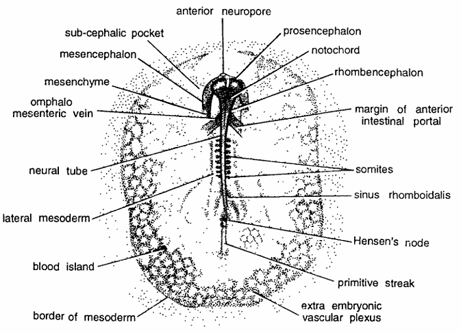
- Entire embryo of 28 hours of incubation shows marked advance in the development of cephalic region.
- Neural folds completely fuse forming neural tube, which becomes completely separated from superficial ectoderm.
- Head projects free from blastoderm.
- Three primary brain vesicles, namely prosencephalon (forebrain), mesencephalon (mid-brain) and rhombencephalon (hind brain) are differentiated.
- Eight pairs of somites develop.
- Anterior neuropore still remains open.
- Hensen’s node still pushed back and primitive streak becomes much smaller.
- Other structures seen in section are anterior intestinal portal, extra-embryonic vascular plexus, blood island, omphalomesenteric vein and sub-cephalic pocket, blood island, lateral mesoderm border of mesoderm and mesenchyme
Identification : Since the embryo shows divisions of brain cavities and above features, hence it is whole mount of chick embryo of 28 hours of incubation.
Chick Embryo : T.S. Passing Through 28 Hours of Incubation
Comments
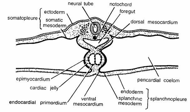
- T.S. through 28 hours of incubation shows formation of heart from mesoderm
- Epi-myocardium, endocardial primordium and cardiac jelly are seen.
- The section also shows closed neural canal, notochord, line of fusion of margins of anterior intestinal portal, somatopleure, splanchnopleure, pericardial coelom, foregut, dorsal mesocardium and ventral mesocardium are seen.
Identification : Since the section contains endocardial primordium and above structure hence it is T.S. of chick embryo of 28 hours of incubation
Chick Embryo : Whole Mount of 33 Hours of Incubation
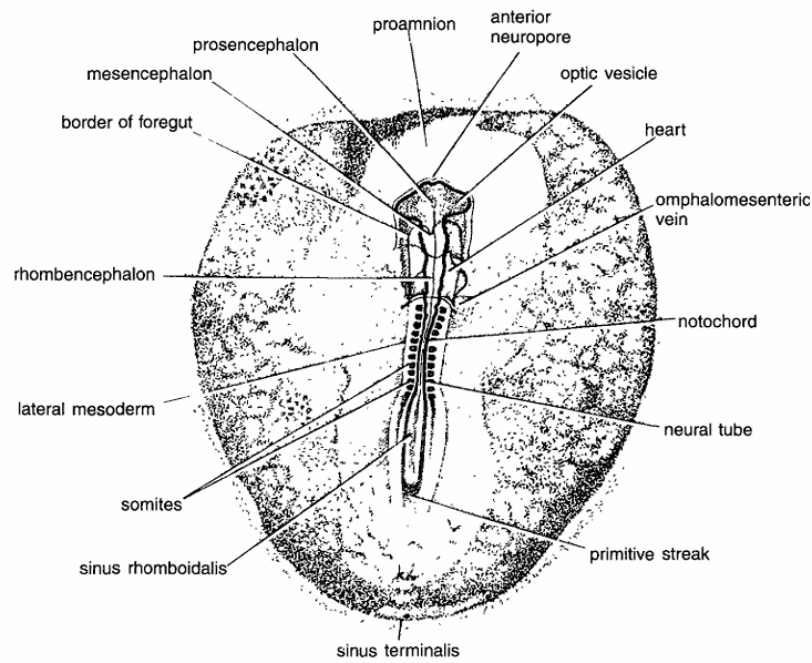
Comments
- Thirty-three hours incubated embryo shows some of the fundamental steps in the formation of central nervous system and circulatory system.
- Various neuromeric enlargements form brain regions. Brain is differentiated into prosencephalon (fore brain), mesencephalon (mid brain) and rhombencephalon (hind brain).
- Optic vesicles are established as paired lateral outgrowths of the prosencephalon.
- Infundibulum is formed as a sort of depression in the floor of the prosencephalon.
- Twelve pairs of somites are formed. Mid-region of the heart is considerably dilated and bent to the right.
- Anterior omphalomesenteric veins have developed. Border of foregut is formed.
- Primitive streak becomes shorter because of the lengthening of the neural tube.
- Proaminon, neural tube, notochord, sinus rhomboidalis and sinus terminalis are also present.
Identification : Since it has 12 pairs of somites and above feature hence it is whole mount of chick embryo of 33 hours of incubation.
Chick Embryo : T.S. Passing Through 33 Hours of Incubation
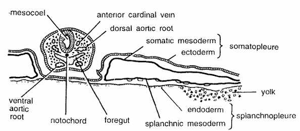
Comments
- This section shows ectoderm, prosocoel, opticoel, mesenchyme, somatic mesoderm, splanchnic mesoderm and endoderm and yolk
- It shows mid-structures namely, mesocoel, anterior cardiual vein, dorsal aortic root, somatopleure, splanchnopleure, foregut, notochord and ventral aortic root.
Identification : Since the section shows somatopleure, splanchnopleure and above features hence it is T.S. of chick embryo passing through 33 hours of incubation
Chick Embryo : Whole Mount of 43 Hours of Incubation
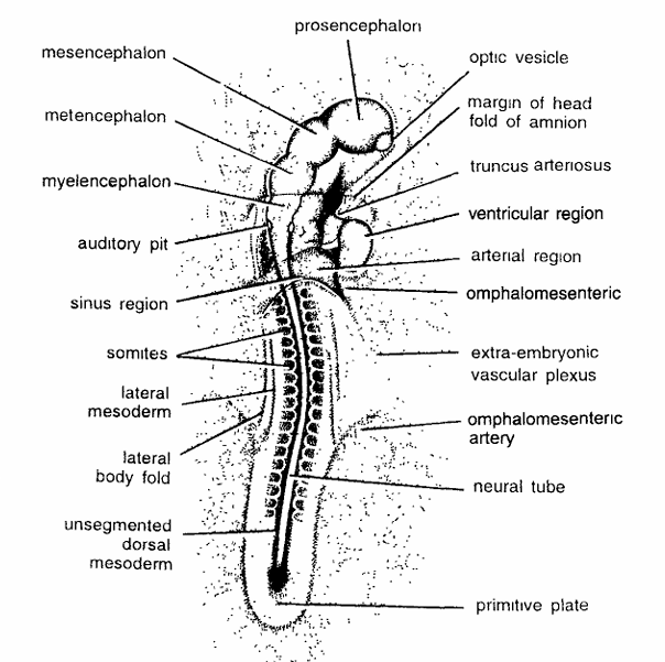
Comments
- 43 hours incubated embryo shows cranial flexure and torsion.
- Cephalic region of embryo is twisted in such a way that left side comes to lie next to the yolk and right side away from yolk.
- Brain is differentiated into prosencephalon, mesencephalon, metencephalon and myelencephalon.
- Heart becomes 4 chambered.
- It is differentiated into ventricular, arterial and sinus regions. Truncus arteriosus also develops. Auditory pit also makes its appearance.
- Vitelline vessels communicate with omphalomesenteric vein.
- Omphalomesenteric arteries and extra-embryonic vascular plexus are well developed.
- Nineteen pairs of somites are formed. Primitive streak diminishes to a small primitive plate.
- Head fold margin develops. Neural plate becomes well developed.
- Lateral mesoderm insegmented mesoderm is seen.
- After 43 hours of incubation, the embryo develops rudiments of most of the organs.
Identification : : Since it has 10 pairs of somites and above features, hence it is whole mount of chick embryo of 43 hours of incubation.
Chick Embryo : Whole Mount of 72 Hours of Incubation
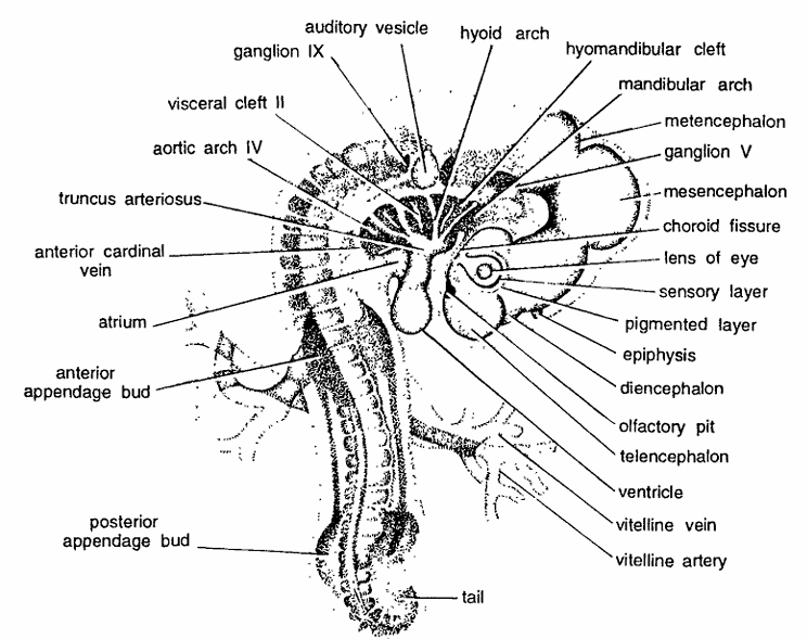
Comments
- After 72 hours of incubation, body is affected throughout by torsion and entire body is turn to 90°.
- Torsion is complete, well posterior to the level of heart, but caudal portion of embryo is not turned on its side.
- Due to the cranial and cervical flexures, the long axis of the embryo shows nearly right angled bends in the mid brain and in the neck region.
- Mid body becomes concave.
- Visceral arches develop.
- Mandibular arch forms caudal boundary of oral depression and becomes more distinct.
- Nasal pits appear as shallow depressions.
- Appendage rudiments also make their appearance.
- Cephalization is going on. Telencephalon also develops anterior appendage bud near mid body and posterior appendage near the tail.
- In the eye, lens, sensory and pigmented layers are differentiated.
- Number of somites increases to 36 pairs. Vitelline arteries and vitelline veins also make their appearance.
Identification : Since it contains 36 pairs of somites and above features, hence it is whole mount of chick
embryo of 72 hours of incubation.
DEVELOPMENT OF CHICK EMBRYO : PRESERVED
Take one dozen fertilized eggs and incubate them is a incubator. Take out incubated eggs after 3, 6, 12, 18 and 21 days and preserved them in formaline. Study the developing embryos.
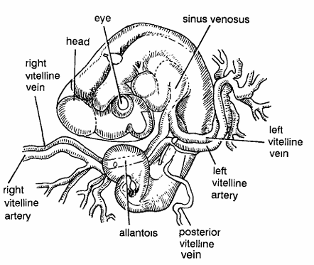
Chick Embryo : Three Days Old Formalin Preserved Whole Embryo from Ventral Side
Comments
- After 3 days to embryo after torsion becomes like telephone receiver. It bends at 90 degree.
- Body divided into broad head and narrow tail.
- Three divisions of brain namely prosencephalon, mesencephalon and metancephalon are clearly seen.
- Optic and auditory capsules have developed. Sinus venosus has started developing.
- Vitelline and allantoic vessels have started appearing. Right vitelline artery, posterior vitelline artery, left vitelline artery and left vitelline vein have appeared.
Identification : Since the embryo has shape of telephone receiver and above features, hence it is chick embryo 3 days old.
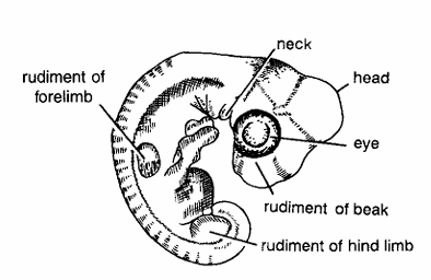
Chick Embryo : 6 Days Old Formalin Preserved Whole Embryo from Dorsal Side
Comments
- After 6 days embryo has considerably grown with broad head and narrow bluntly ending tail.
- Prosencephalon, mesencephalon and metencephalon have distinctly developed.
- Optic vesicle has developed more.
- Rudiments of anterior and posterior limbs are seen.
- Toes contain claw.
- Near head beak is also appearing.
Identification : Since above specimen has beak rudiment and above features, hence it is chick embryo 6 days old.
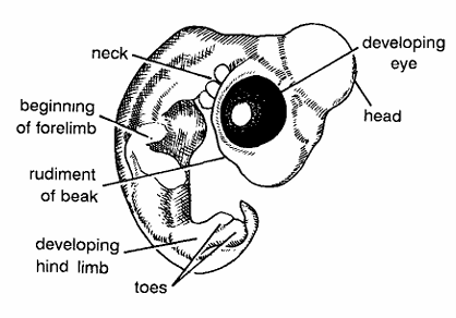
Chick Embryo : 12 Days Old Formalin Preserved Whole Embryo from Dorsal Side
Comments
- After 12 days chick embryo take the shape of a bird.
- Body is distinctly divided into head, neck, trunk and tail.
- Head contains eyes with developing eyelids.
- Upper and lower beaks develop.
- Forelimbs develop into wings.
- Hind limbs develop clawed toes
Identification : Since above specimen has beak rudiment and above features, hence it is chick embryo 6 days old.
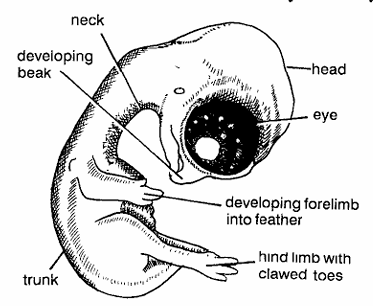
Chick Embryo 18 Days Old Formalin Preserved Whole Embryo from Dorsolateral Side
Comments
- At this stage young chick considerably acquires features of adult bird.
- Body divided into head, neck, trunk and tail.
- Eyes become well developed along with eyelids.
- Pointed beak develops like pig. Entire body is covered with down feathers called as plumes.
- Wings develops.
- Hind limb develop claw.
Identification : Since the specimen contains down feathers and above features, hence it is 18 days old chick embryo.
Chick Embryo 21 Days Just Hatched Chick
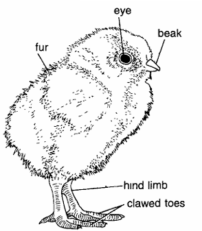
Comments
- After 21 days chick comes out of the egg.
- Except beak and toes, entire body is covered with soft whitish down feathers which are called as plumes.
- Plumes are softer than silk and they do not contain rachis.
- Plumes form natal covering over young chick.
- Body is divided into head, neck, trunk and tail.
- Eyes and beak well developed.
- Rudiments of developing flying feathers distinct.
Identification : Since the chick contains over, feather all over body except beak and claws hence it is just hatched chick.
