
Herdmania Slides : Metamorphosis : Tadpole Larva
Larva of Herdmania is called as tadpole larva. After hatching it leads a free-swimming life for 90-180 seconds and then undergoes retrogressive metamorphosis.
Comments :
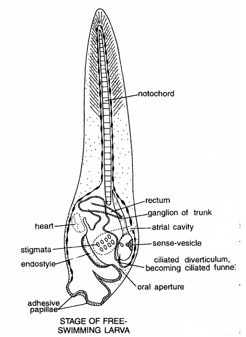
- Tadpole larva
- Larva is minute, transparent and 1.2 mm long. Body divisible into head or trunk and a long posterior tail
- Anteriorly trunk contains 2 dorsolateral and 1 ventral median adhesive papillae.
- Tail contains tail fin which is extension of thin covering of the body.
- Along tail fin striations are present giving impression as fin rays.
- In the axis of tail notochord is present. Structures seen adhesive papillae, stigmata, heart rectum, ganglia of trunk, atrium on ciliated funnel.
- Various internal organs comprise sensory vesicle with otolith, visceral ganglion, mouth, pharynx, endostyle, stomach, intestine, atrium and atriopore.
- Tail is a powerful locomotory organ. It has notochord, nerve cord and tail fin.
2. Metamorphosis
During metamorphosis, tadpole larva undergoes both retrogressive and progressive changes. Retrogressive changes : The just hatched tadpole larva immediately attaches to substratum by adhesive papillae. It undergoes following degenerative changes
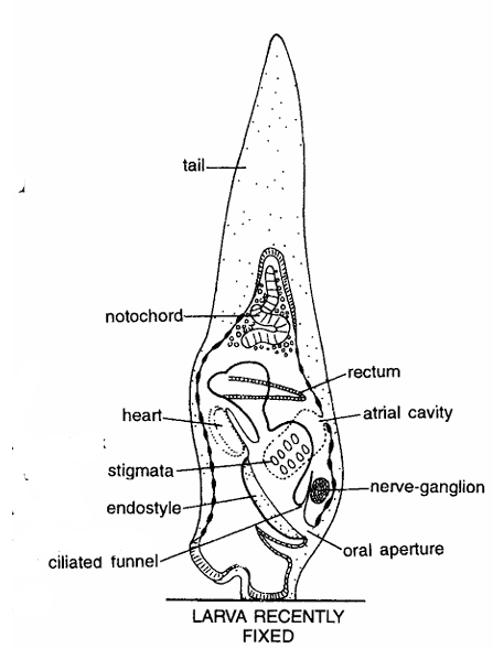
- Larval tail with tail fin shortens and later on disappears along with notochord and nerve cord.
- Anterior region expands. Sense organs and adhesive papillae disappear.
- Other structures seen are rectum, atrium, nerve ganglion, ciliated diverticulum, oral aperture, endostyle, stigmata or gill slits, heart and notochord.
3. Progressive changes
Larva undergoes progressive changes. Young adult becomes without tail. Branchial and atrial apertures become functional, pharynx develops gill slits. Young adult undergoes following progressive changes
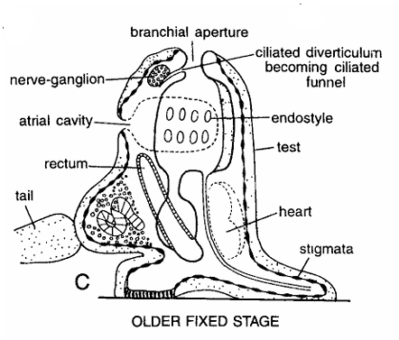
- Body becomes surrounded by test or tunic all over. Tail completely degenerates. Branchial and atrial apertures become functional.
- Pharynx enlarges and develops gill slits. Stomach, intestine and liver develop.
- Heart, pericardium and gonads appear.
- Other structures seen are nerve ganglion, neural duct, endostyle, dorsal tubercle, atrial cavity, stolon, ciliated groove, atrium, and rectum.
- Special features : All chordate characters are found only in tadpole larva of Herdmania. Adults are without any chordate characters. Larva shows unique combination of retrogressive and progressive metamorphosis.
Herdmania Slides : Neural Complex
Comments
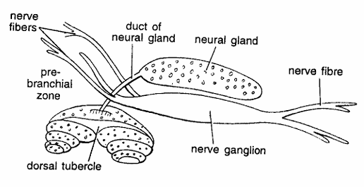
- Nervous system, excretory system and associated receptor organs are together called as neural complex.
- It is composed of neural gland, nerve ganglion and dorsal tubercle.
- Neural gland is oval-shaped structure, found in the mantle between two siphons and just above the nerve ganglion.
- Anteriorly, it gives a called an duct of neural gland duct, which opens by a ciliated funnel near the base of dorsal tubercle.
- It contains central tubule, which gives off a few branching peripheral tubules.
- Neural gland is supposed to be excretory in function.
- It secretes excretory material appearing as pigmented granules in the substance of the gland.
- Cells impregnated with such granules are discharged into the branchial sac.
- Posteriorly neural gland given rise to nerve fibres.
- Dorsal ganglion or nerve ganglion is solid, elongated structure, found dorsally nerve between branchial and atrial openings. It fibers constitutes central nervous system. It gives 3 branches to branchial siphon and 2 nerves to atrial siphon.
- Experimental removal of brain indicates that it is not very much needed for the life and metabolic activities.
- Dorsal tubercle has a broad base from which 2 spirally coiled cones originate. Each cone has 3 coils of spirally ciliated channel. It is supposed to be chemosensory.
Identification : Since it contains dorsal tubercle and above features, hence it is neural complex of Herdmania.
Herdmania Slides : Various Spicules
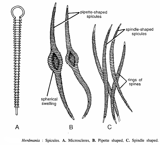
Comments
Spicules of Herdmania are found embedded in test, body wall and organ systems and are calcareous in nature. Spicules are of three kinds.
- Microscleres : These are embedded in test, are microscopic and measure 40 to 80 microns long. Each spicule has a small knob-like head and an elongated body containing several spiny rings.
- Pipette-shaped : Found in body wall, particularly in the region of gonads and liver. They are swollen in middle, pointed at tips and measure 3.5 mm in length.
- Spindles shaped : Usually found associated with blood vessels and part of alimentary canal, measure 1.5 to 2.5 mm in length and contain 15 to 20 rings of minute spines.
Special features : Spicules protect the animal from predators. They serve to attach body wall with the test. They also form the framework of certain passages for blood vessels.
