
The antibody structure is closely related to its biological function. A typical immunoglobulin G (IgG) molecule has a characteristic Y-shaped arrangement consisting of two identical heavy chains and two identical light chains. These polypeptide chains are connected by disulfide bonds and stabilized by additional molecular interactions. The two arms contain antigen-binding sites, while the stem participates in interactions with immune cells and other effector molecules.
Antibodies are specialized proteins of the adaptive immune system that recognize specific molecular structures called antigens. They are also known as immunoglobulins (Ig) and are produced in large quantities by plasma cells, which develop from activated B lymphocytes. These molecules help protect the body against pathogens, toxins and other potentially harmful substances through specific antigen recognition and several immune-defense mechanisms.
An antibody molecule contains both variable and constant regions. The variable regions differ among antibodies and form the antigen-binding sites, allowing different antibodies to recognize different epitopes. In contrast, the constant regions are relatively conserved within an immunoglobulin class and contribute to the molecule’s biological properties. This arrangement allows antigen recognition to be linked with appropriate immune responses.
The major structural regions include the Fab region, Fc region, hinge region, variable domains and constant domains. Each has a distinct role. The Fab portions recognize and bind antigens, the Fc portion mediates many effector functions, and the hinge region provides flexibility in immunoglobulins that possess a conventional hinge. The detailed organization of these components helps explain how a single molecule can combine specific recognition with immune activity.
Although IgG provides the most commonly used model for studying antibody structure, not all immunoglobulins have identical arrangements. IgM may form a pentamer when secreted, secretory IgA commonly occurs as a dimer, and IgE and IgD are generally monomeric. These differences influence their distribution, binding properties and biological roles.
Understanding the molecular organization of antibodies is essential for studying antigen recognition, antibody diversity, class switching, affinity maturation and humoral immunity. It also provides the foundation for understanding diagnostic techniques and therapeutic monoclonal antibodies. For a general overview of the five immunoglobulin classes and their roles, see our related article, Antibody: Definition, Structure, Types and Functions.
Basic Antibody Structure
A typical IgG antibody is a Y-shaped glycoprotein composed of four polypeptide chains: two identical heavy chains and two identical light chains. Each chain contains immunoglobulin domains that fold into compact structures stabilized mainly by intrachain disulfide bonds and other non-covalent interactions. The chains associate to form a functional molecule with two antigen-binding sites.
The Y-shaped arrangement can be divided into two arms and one stem. The arms contain the antigen-binding portions, while the stem forms the principal Fc region. A flexible hinge connects the arms to the stem in IgG, allowing the binding sites to adopt different orientations when encountering antigens.
2.1 Four Polypeptide Chains
The two heavy chains are longer and contribute most of the molecule’s central framework and Fc region. Each heavy chain also contributes a variable domain to one antigen-binding site.
The two light chains are shorter. Each light chain associates with one heavy chain in an arm of the molecule. The variable domains of a heavy chain and its associated light chain work together to form an antigen-binding site.
The four chains are arranged symmetrically in a typical IgG molecule. This organization gives the antibody two equivalent antigen-binding sites, although their effective binding can depend on the antigen’s structure and accessibility.
Antigen-Binding Arms
Each arm contains the variable domains of one heavy chain and one light chain, followed by constant domains. The variable domains are designated (V_H) and (V_L). Their combined surfaces form the antigen-binding site, which recognizes a particular epitope through molecular complementarity.
The two arms allow a typical IgG antibody to bind two epitopes. These may be on separate antigen molecules or on repeated sites within a larger antigenic structure. The ability to bind more than one site can promote cross-linking of antigens, although the resulting biological effect depends on the antigen and experimental conditions.
Stem or Fc Portion
The stem is formed mainly by the constant domains of the two heavy chains. In IgG, this region includes the (C_H2) and (C_H3) domains from each heavy chain. The Fc portion does not provide the conventional antigen-binding sites; instead, it interacts with Fc receptors and, for appropriate antibody subclasses and complexes, complement components.
These interactions help connect antibody binding to downstream activities such as phagocytosis, cellular activation and complement-mediated defense.
Disulfide Bonds and Molecular Stability
Disulfide bonds form between cysteine residues and help maintain the integrity of the antibody molecule. Intrachain disulfide bonds stabilize individual immunoglobulin domains, while interchain disulfide bonds link heavy and light chains and connect the heavy chains to each other.
The exact number and arrangement of these bonds vary among immunoglobulin classes and subclasses. Together with non-covalent interactions, they help maintain the three-dimensional structure required for antigen recognition and effector activity.
Main Structural Components
| Component | Structural location | Main role |
|---|---|---|
| Heavy chains | Form the central framework, arms and stem | Contribute to antigen binding and determine immunoglobulin class through their constant regions |
| Light chains | Associate with heavy chains in the arms | Contribute to antigen-binding sites |
| Variable domains | Tips of the two arms | Provide antigen-recognition specificity |
| Constant domains | Follow variable domains and form much of the stem | Support structure and contribute to effector functions |
| Fab regions | Two antigen-binding arms | Recognize and bind epitopes |
| Fc region | Stem of the molecule | Interacts with Fc receptors and certain complement components |
| Hinge region | Between arms and stem in IgG | Provides flexibility |
| Disulfide bonds | Within and between chains | Stabilize domains and connect polypeptide chains |
The basic arrangement of an IgG molecule provides the framework for understanding the more specific roles of heavy chains, light chains, variable regions, constant regions and antigen-binding sites, which are discussed in the following sections.
Heavy Chains
Heavy chains are the longer polypeptide chains of an antibody molecule. A typical IgG antibody contains two identical heavy chains, each associated with one light chain. Together, the heavy chains contribute to the two antigen-binding arms and form most of the central stem, including the Fc region.
Each heavy chain consists of one variable domain, designated (V_H), followed by constant domains designated (C_H1), (C_H2) and (C_H3) in IgG. These domains fold into compact immunoglobulin structures. Their arrangement supports the stability of the molecule and allows the variable and constant portions to perform different functions.
Variable Domain of the Heavy Chain
The variable heavy-chain domain ((V_H)) lies near the tip of each antibody arm. It associates with the variable domain of a light chain ((V_L)) to form an antigen-binding site.
The amino-acid sequence of (V_H) differs among antibodies. Within this domain are three complementarity-determining regions (CDR1, CDR2 and CDR3), interspersed with framework regions. The CDRs form much of the antigen-contacting surface, while the framework regions maintain the domain’s three-dimensional structure.
The heavy-chain CDR3 is often especially diverse because its sequence is generated through the joining of gene segments during B-cell development, together with additional junctional diversity. This diversity contributes greatly to the range of antigens that the immune system can recognize.
Constant Domains of the Heavy Chain
In IgG, the constant portion of each heavy chain includes three domains:
- (C_H1): Located in the Fab portion, where it associates with the light-chain constant domain and helps stabilize the antibody arm.
- (C_H2): Part of the Fc region; in IgG, it contains an important site of N-linked glycosylation that influences Fc structure and interactions.
- (C_H3): Also part of the Fc region, contributing to its stability and interactions with other molecular components.
The organization differs among immunoglobulin classes. IgM and IgE heavy chains have four constant domains and lack the conventional flexible hinge found in IgG. IgA, IgD and IgG have a hinge region, although its length and sequence vary. These differences affect molecular flexibility and biological properties.
Heavy Chains Determine the Immunoglobulin Class
The constant region of the heavy chain determines the immunoglobulin isotype. The five major human immunoglobulin classes use the following heavy-chain types:
| Immunoglobulin class | Heavy-chain type |
|---|---|
| IgG | Gamma ((\gamma)) |
| IgM | Mu ((\mu)) |
| IgA | Alpha ((\alpha)) |
| IgE | Epsilon ((\varepsilon)) |
| IgD | Delta ((\delta)) |
The variable region determines antigen specificity, whereas the heavy-chain constant region contributes to the antibody’s class-specific properties and effector functions. For example, IgG can interact with Fc gamma receptors, while IgE binds with high affinity to Fc epsilon RI receptors on mast cells and basophils.
3.4 Functions of Heavy Chains
Heavy chains contribute to antibody activity in several ways:
- Antigen recognition: Their variable domains contribute directly to the antigen-binding sites.
- Structural stability: Interactions with light chains and other heavy-chain regions maintain the molecule’s organization.
- Effector activity: Constant domains interact with Fc receptors and, in appropriate circumstances, complement components.
- Class-specific properties: Heavy-chain type influences antibody distribution, molecular form and biological activity.
- Molecular flexibility: Hinge regions in applicable classes allow the arms to change orientation relative to the Fc portion.
Thus, heavy chains are not merely structural supports. They contribute to antigen recognition and determine many of the biological properties that distinguish one immunoglobulin class from another.
Light Chains
Light chains are the shorter polypeptide chains of an antibody. A typical immunoglobulin molecule contains two identical light chains, each paired with one heavy chain. Although shorter than heavy chains, light chains are essential components of the antigen-binding sites.
There are two types of immunoglobulin light chains in humans: kappa ((\kappa)) and lambda ((\lambda)). An individual antibody normally contains either two kappa light chains or two lambda light chains. It does not ordinarily contain one of each type.
Structure of a Light Chain
Each light chain contains two immunoglobulin domains:
- Variable light-chain domain ((V_L))
- Constant light-chain domain ((C_L))
The (V_L) domain is located near the tip of the antibody arm and pairs with the (V_H) domain of the associated heavy chain. Together, these two variable domains form the antigen-binding site.
The (C_L) domain lies closer to the central part of the arm. It associates with the (C_H1) domain of the heavy chain, helping maintain the structure of the Fab region.
Both light-chain domains are stabilized by intrachain disulfide bonds and other molecular interactions that support their folded structures.
Variable Region of the Light Chain
The variable light-chain domain ((V_L)) contributes to the antigen-binding surface. Like (V_H), it contains three complementarity-determining regions—CDR1, CDR2 and CDR3—separated by framework regions.
The CDRs of the heavy and light chains work together to create a binding surface whose shape and chemical properties complement a particular epitope. Some antibodies rely more heavily on heavy-chain contacts, while others use substantial contributions from both chains.
Changes in the amino-acid sequence of the variable domains can alter antigen specificity and affinity. The combination of different heavy- and light-chain variable regions therefore contributes to antibody diversity.
Constant Region of the Light Chain
The constant light-chain domain ((C_L)) is more conserved than the variable domain within a particular light-chain type. It provides structural support and interacts with the heavy chain in the Fab portion.
Unlike the heavy-chain constant region, the light-chain constant region does not determine whether an antibody belongs to IgG, IgM, IgA, IgE or IgD. That classification depends on the heavy-chain constant region.
Kappa and lambda light chains can both occur in antibodies of any of the five major immunoglobulin classes.
Kappa and Lambda Light Chains
Kappa and lambda light chains differ in their constant-region sequences and in the gene segments used during their development. Both types can support antigen recognition and participate in the formation of functional antibodies.
The presence of kappa or lambda chains does not, by itself, define antigen specificity or immunoglobulin class. Specificity depends primarily on the combined variable regions, while the heavy-chain constant region defines the isotype.
Laboratory measurement of free kappa and lambda light chains can be useful in evaluating certain plasma-cell disorders, because abnormal production may change their concentrations or ratio. This is a clinical application of light-chain biology rather than a separate antibody class.
Functions of Light Chains
Light chains perform several essential roles:
- Contribute to antigen binding: The (V_L) domain forms part of each antigen-binding site.
- Support structural stability: The (C_L) domain associates with the heavy chain in the Fab region.
- Contribute to binding specificity: Light-chain CDRs help determine how the antibody recognizes its epitope.
- Support antibody diversity: Different variable light-chain sequences combine with heavy-chain variable regions to generate a broad range of antigen-recognition sites.
Heavy Chains vs Light Chains
| Feature | Heavy chains | Light chains |
|---|---|---|
| Number in a typical antibody | Two | Two |
| Relative length | Longer | Shorter |
| Domains in IgG | One variable and three constant domains | One variable and one constant domain |
| Variable domain | (V_H) | (V_L) |
| Constant domains | (C_H1), (C_H2), (C_H3) in IgG | (C_L) |
| Contribution to antigen binding | Provides one side of each binding site | Provides the other side of each binding site |
| Determines immunoglobulin class | Yes | No |
| Main types | Gamma, mu, alpha, epsilon and delta | Kappa and lambda |
In summary, the heavy and light chains work as an integrated molecular unit. Their variable domains form the antigen-binding surface, while their constant domains and interchain associations maintain the structure required for antibody recognition and activity.
Variable and Constant Regions
An antibody contains variable and constant regions that differ in amino-acid sequence, structural organization and biological role. The variable regions enable specific antigen recognition, whereas the constant regions provide structural support and determine many of the antibody’s effector properties.
Both heavy and light chains contain a variable domain near the amino-terminal end. In a typical IgG molecule, each heavy chain has one variable domain and three constant domains, while each light chain has one variable domain and one constant domain. The association of these domains produces the functional architecture of the antibody.
Variable Region
The variable region is located at the tips of the two arms of a typical antibody. It consists of the variable heavy-chain domain ((V_H)) and variable light-chain domain ((V_L)), which come together to form an antigen-binding site.
The amino-acid sequences of these domains differ among antibodies. This variation arises through genetic mechanisms during B-cell development and can be further modified in activated B cells through somatic hypermutation. Consequently, the immune system can produce a vast repertoire of antibodies with different antigen specificities.
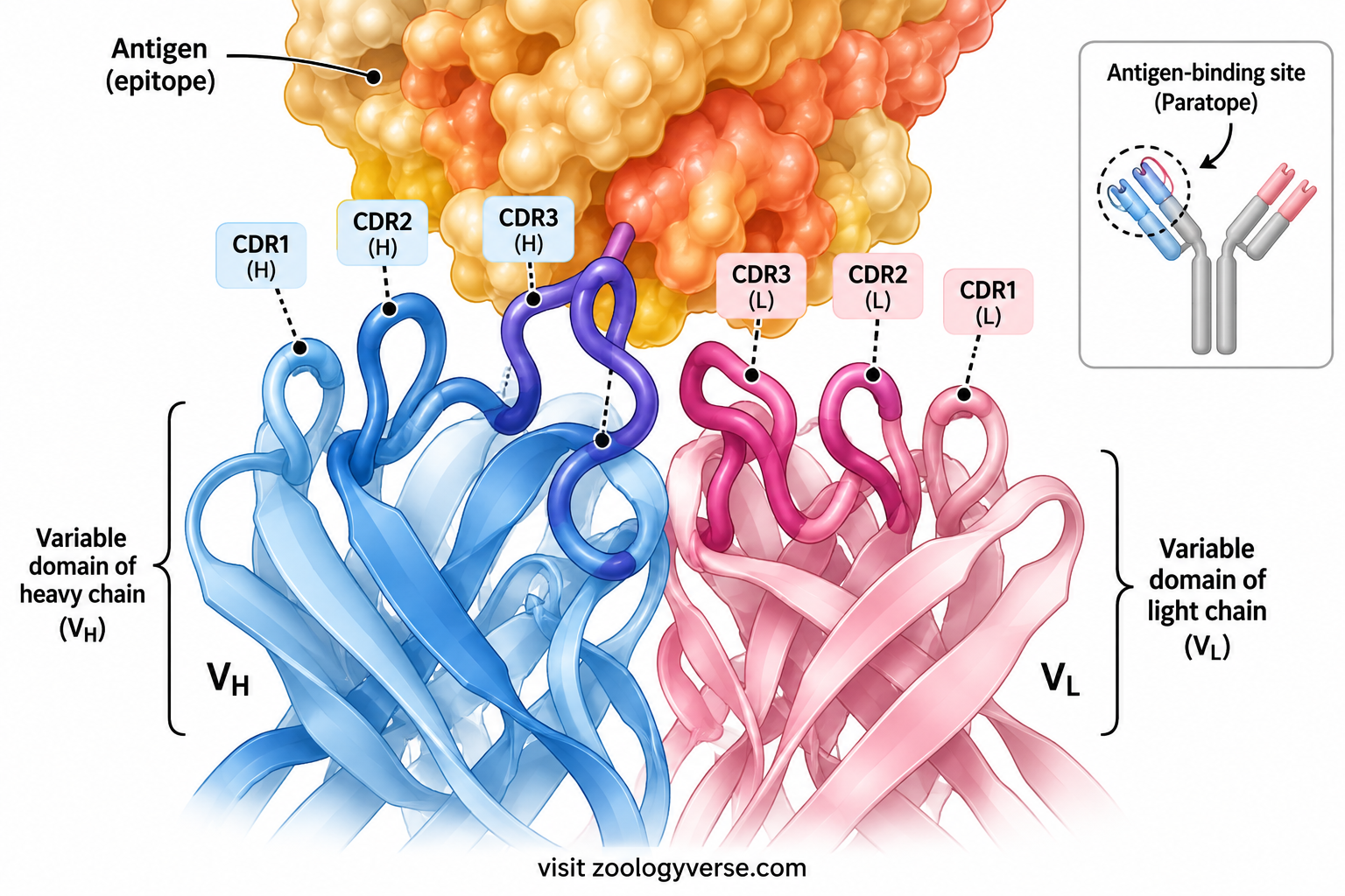
Within each variable domain are three complementarity-determining regions (CDRs). These loops are particularly variable and contribute much of the surface that contacts the antigen. The remaining portions, called framework regions, support the correct folding and positioning of the CDRs.
The variable region does not usually recognize every part of an antigen equally. Instead, it binds a particular molecular site called an epitope. Recognition depends on the shape, charge distribution, hydrogen-bonding potential and other chemical properties of the binding surface.
Constant Region
The constant region has a comparatively conserved amino-acid sequence within a given immunoglobulin class or subclass. It is found after the variable domain in each chain and contributes to the structural stability of the molecule.
The heavy-chain constant region is especially important because it determines the immunoglobulin class—IgG, IgM, IgA, IgE or IgD. Different classes interact with different receptors, have different distributions in the body and perform specialized immune functions.
For example, the Fc region of appropriate IgG subclasses can bind Fc gamma receptors on immune cells, while IgE binds with high affinity to Fc epsilon RI receptors on mast cells and basophils. The constant region of IgA supports its role in mucosal defense, and secretory IgA has an additional secretory component acquired during epithelial transport.
The light-chain constant region, designated (C_L), contributes mainly to the organization and stability of the Fab region. It does not determine the antibody’s immunoglobulin class.
Framework Regions and Complementarity-Determining Regions
The variable domains contain both framework regions and CDRs.
- Framework regions: These relatively conserved segments form the structural scaffold of each variable domain.
- Complementarity-determining regions: These variable loops contribute directly to antigen recognition and contact with epitopes.
The three CDRs of a heavy-chain variable domain and the three CDRs of a light-chain variable domain together form the principal antigen-contacting surface in a conventional antibody. Their combined geometry helps determine which epitopes can be recognized.
Variable Region vs Constant Region
| Feature | Variable region | Constant region |
|---|---|---|
| Sequence | Highly variable among antibodies | More conserved within an isotype or subclass |
| Main location | Near the tips of the arms | Follows the variable domains and contributes to the rest of the molecule |
| Heavy-chain designation | (V_H) | (C_H) domains |
| Light-chain designation | (V_L) | (C_L) |
| Primary role | Antigen recognition and specificity | Structural support and, particularly for heavy chains, effector functions |
| CDRs | Present | Absent |
| Determines immunoglobulin class | No | Heavy-chain constant region does |
| Example | Different antibodies recognize different epitopes | IgG and IgA have different heavy-chain constant regions |
The distinction between these regions explains how antibodies can recognize an enormous variety of targets while using a broadly conserved molecular framework.
Antigen-Binding Sites
An antigen-binding site is the molecular surface of an antibody that specifically recognizes an epitope on an antigen. A typical IgG molecule has two antigen-binding sites, one at the tip of each arm.
Each site is formed by the paired variable domains of one heavy chain and one light chain. The heavy-chain variable domain ((V_H)) and light-chain variable domain ((V_L)) contribute complementary surfaces that together determine how the antibody interacts with its target.
Formation of the Antigen-Binding Site
The antigen-binding site develops from the folding and association of the (V_H) and (V_L) domains. The three-dimensional arrangement of their CDR loops creates a surface that can accommodate a particular epitope.
The interaction is based on molecular complementarity rather than a rigid lock-and-key fit. Both the antibody and antigen have three-dimensional structures, and their surfaces may adjust slightly during binding.
The antigen-binding site can recognize epitopes located on proteins, carbohydrates and other molecular structures, depending on the antibody’s specificity. However, recognition differs between B-cell receptors and T-cell receptors: conventional antibodies and B-cell receptors can bind accessible epitopes on intact antigens, whereas conventional T-cell receptors generally recognize antigen-derived peptides presented by major histocompatibility complex (MHC) molecules.
For more detail on the target molecules themselves, see Antigen: Definition, Types, Structure and Functions.
Epitope Recognition
An epitope, also called an antigenic determinant, is the specific part of an antigen recognized by an antibody or another antigen receptor.
A single antigen may contain multiple epitopes. Different antibodies can recognize different sites on the same antigen, and a polyclonal antibody preparation may contain several antibody populations directed against different epitopes.
Epitope accessibility is important. A potential binding site may be hidden within a folded protein, covered by another molecule or altered by chemical modification. Therefore, the presence of an epitope does not automatically mean that every antibody directed against it can bind under all conditions.
Forces Involved in Antigen Binding
Antigen-antibody binding is generally mediated by several types of non-covalent interactions:
- Hydrogen bonds: Form between suitable hydrogen-bond donors and acceptors.
- Electrostatic interactions: Arise between charged or partially charged groups.
- Hydrophobic interactions: Favor the association of compatible non-polar surfaces in an aqueous environment.
- Van der Waals forces: Contribute through close-range interactions between atoms.
No single interaction necessarily explains the entire binding event. The combined geometry and chemical compatibility of the interacting surfaces determine the overall stability and specificity of the complex.
Specificity and Affinity
Specificity describes an antibody’s preference for a particular epitope or a structurally related set of epitopes. It depends mainly on the arrangement and chemical properties of the variable regions.
Affinity refers to the strength of binding between one antigen-binding site and one epitope. Higher affinity generally means that the complex is more stable under comparable conditions.
Affinity should not be confused with avidity, which describes the combined strength of multiple binding interactions. A multivalent antibody may have high avidity because it can form several simultaneous interactions, even when the affinity of an individual binding site is moderate.
Why Antibodies Have Two Antigen-Binding Sites
The two binding sites of a typical IgG molecule can engage two accessible epitopes. This can increase the overall stability of binding and allow antigens to be cross-linked.
However, effective binding depends on the distance and orientation between epitopes, the flexibility of the antibody and the physical arrangement of the antigen. Having two binding sites does not guarantee that both will always be occupied at the same time.
The two-site arrangement is therefore an important structural feature, but its functional advantage depends on the biological context.
Main Features of an Antigen-Binding Site
| Feature | Explanation |
|---|---|
| Location | At the tips of the antibody arms |
| Main location | One (V_H) and one (V_L) domain |
| Key sequences | Six CDR loops across the two variable domains |
| Target | An epitope on an antigen |
| Binding forces | Non-covalent molecular interactions |
| Specificity | Determined by variable-region structure and chemistry |
| Number in typical IgG | Two |
| Functional outcome | Specific recognition that may lead to neutralization, cross-linking or other immune effects |
The antigen-binding site is the central recognition element of an antibody. Its structure allows the immune system to distinguish among molecular targets, while the remaining regions of the molecule help translate recognition into an appropriate immune response.
Complementarity-Determining Regions (CDRs)
Complementarity-determining regions (CDRs) are short, highly variable loops within the variable domains of an antibody. They contribute directly to the molecular surface that recognizes and binds an antigenic epitope. Their sequences and three-dimensional arrangement are major determinants of antibody specificity.
A conventional antibody has three CDRs in its variable heavy-chain domain ((V_H)) and three in its variable light-chain domain ((V_L)). These are designated CDR1, CDR2 and CDR3 in each domain. Together, the six loops form much of the antigen-contacting surface, although the extent to which each loop contributes varies among antibodies.
Location of CDRs
The CDRs are located within the variable domains at the tips of the antibody arms. Each variable domain also contains framework regions, which provide a structural scaffold that holds the CDR loops in their appropriate positions.
The heavy-chain and light-chain variable domains associate with one another. This arrangement brings their CDRs together to form a binding surface capable of recognizing a particular epitope. The resulting surface may contain a pocket, groove, relatively flat region or another shape suited to the target.
CDR1 and CDR2
CDR1 and CDR2 occur in both the heavy-chain and light-chain variable domains. Their amino-acid sequences vary among antibodies, and their shapes contribute to the overall fit between the binding site and an epitope.
These loops may contact different parts of an antigen. Their contribution depends on the particular antibody and target; neither loop has one universal role that applies to every antibody.
CDR3
CDR3 is often particularly diverse, especially in the heavy-chain variable domain. This diversity arises during B-cell development through the joining of gene segments and the addition or removal of nucleotides at their junctions.
Heavy-chain CDR3 spans the junction formed by the variable (V), diversity (D) and joining (J) gene segments. Because the junctional process is highly variable, CDR3 can differ substantially in length and sequence among antibodies. Light-chain CDR3 is generated through the joining of V and J segments and also contributes to antigen recognition.
The diversity of CDR3 is a major source of the immune system’s ability to recognize many different molecular targets. However, antigen specificity depends on the combined structure of the variable domains, not on CDR3 alone.
Role of CDRs in Antigen Binding
CDRs contribute to binding through their three-dimensional shape and chemical properties. Their amino-acid side chains may form hydrogen bonds, electrostatic interactions, hydrophobic contacts and van der Waals interactions with an epitope.
Changes in CDR sequence can alter which antigen is recognized or how strongly it binds. During an immune response, activated B cells may undergo somatic hypermutation in their immunoglobulin variable-region genes. Selection can favor cells producing antibodies with improved affinity for the antigen, a process known as affinity maturation.
CDRs and Antibody Diversity
The diversity of CDRs reflects several biological mechanisms:
- V(D)J recombination: Rearranges immunoglobulin gene segments during B-cell development.
- Junctional diversity: Creates additional sequence variation at gene-segment joining sites.
- Heavy- and light-chain pairing: Combines different variable domains to form a binding site.
- Somatic hypermutation: Introduces mutations into variable-region genes in activated B cells.
- Selection during immune responses: Favors B-cell clones whose antibodies bind the antigen effectively under the conditions of that response.
These mechanisms allow the immune system to produce an extensive repertoire of antigen-binding sites. The resulting diversity is essential for recognizing new pathogens and responding to changing antigenic targets.
CDRs vs Framework Regions
| Feature | CDRs | Framework regions |
|---|---|---|
| Location | Within antibody variable domains | Between and around CDR loops |
| Sequence variation | Generally higher | Generally more conserved |
| Main contribution | Direct antigen contacts and binding specificity | Structural support for variable domains |
| Number per variable domain | Three | Four main framework segments |
| Role in binding | Form much of the antigen-contacting surface | Maintain the arrangement of the binding surface |
CDRs are therefore crucial to the specificity of antibody recognition, while framework regions provide the structural support that allows those loops to function.
Fab Region
The Fab region, short for fragment antigen-binding, is the portion of an antibody responsible primarily for recognizing and binding antigen. A typical IgG molecule has two Fab regions, one in each arm of its Y-shaped structure.
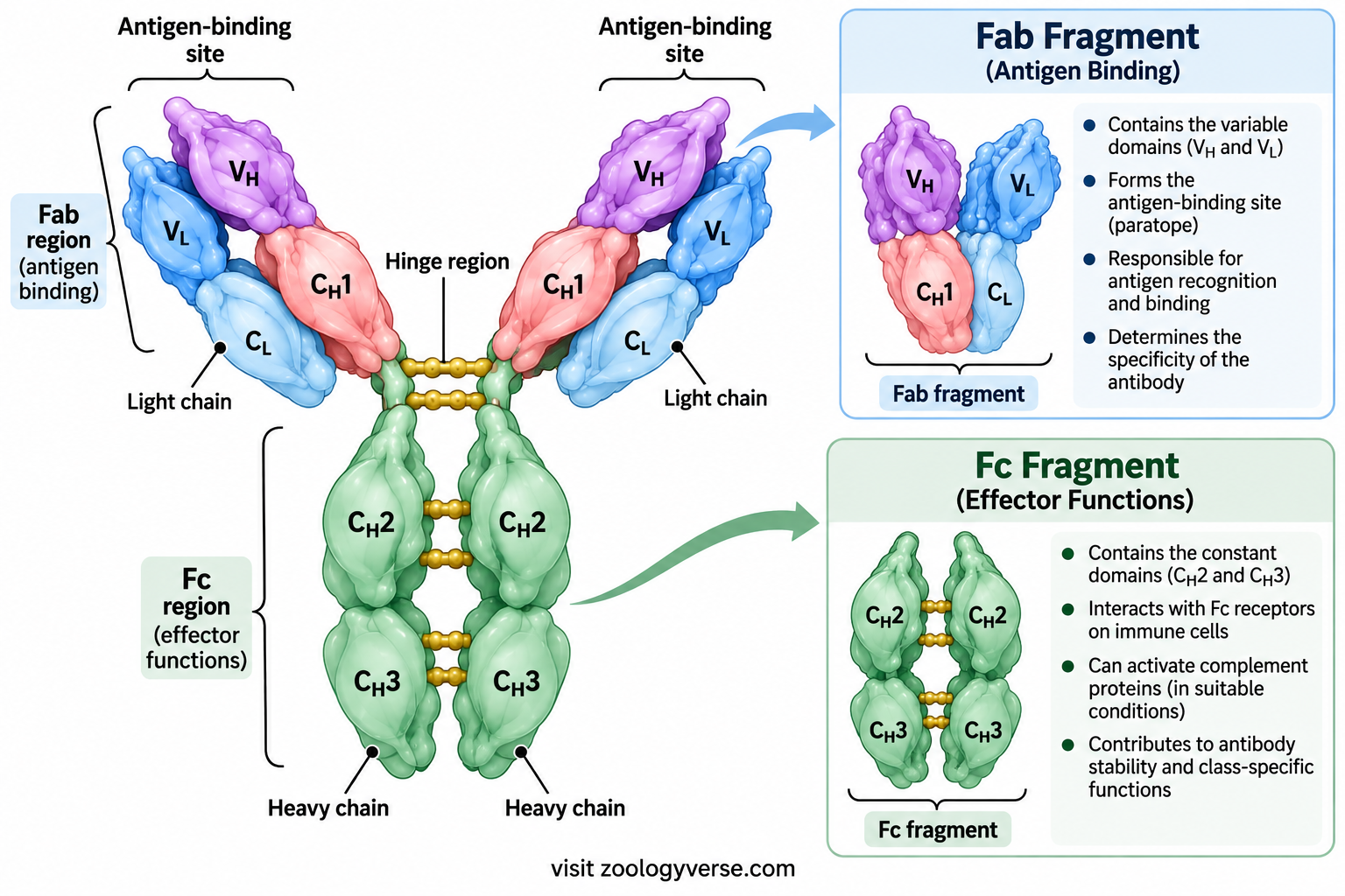
Each Fab region contains one complete light chain and part of one heavy chain. More specifically, it includes the light-chain variable and constant domains ((V_L) and (C_L)) together with the heavy-chain variable and first constant domains ((V_H) and (C_H1)).
This arrangement brings the heavy- and light-chain variable domains together to form the antigen-binding site. The Fab region therefore provides the structural basis for specific antigen recognition.
Structural Organization of Fab
The Fab region contains four immunoglobulin domains:
- Variable heavy-chain domain ((V_H))
- First constant heavy-chain domain ((C_H1))
- Variable light-chain domain ((V_L))
- Constant light-chain domain ((C_L))
The (V_H) and (V_L) domains associate near the tip of the arm, where their CDR loops form the antigen-binding surface. The (C_H1) and (C_L) domains lie closer to the hinge and support the structure of the arm.
Disulfide bonds and non-covalent interactions help maintain the association between the chains and the stability of the Fab region.
Antigen Recognition by Fab
The Fab region recognizes a particular epitope through the complementary structure of its variable domains. Binding depends on the shape, chemical properties and accessibility of the epitope, together with the arrangement of the antibody’s CDR loops.
Although a typical IgG molecule has two Fab regions with the same variable-region specificity, the ability to engage two sites simultaneously depends on the geometry and accessibility of the target. Binding at both sites may increase avidity or promote cross-linking, but this is not guaranteed for every antigen.
Fab and Antibody Specificity
The variable domains within Fab largely determine which epitope an antibody recognizes. Antibodies produced by different B-cell clones may have different CDR sequences and therefore bind different epitopes, even when the epitopes occur on the same antigen molecule.
The Fab region is also important in therapeutic antibody design. Researchers can select or engineer antigen-binding regions to target particular molecules, including proteins involved in infection, cancer or inflammatory disease. However, the overall therapeutic effect also depends on factors such as the antibody’s Fc region, stability, distribution and interaction with the immune system.
Fab vs Fc
The Fab and Fc portions have different but complementary functions. Fab recognizes the antigen, while Fc helps recruit appropriate immune mechanisms after binding.
| Feature | Fab region | Fc region |
|---|---|---|
| Full name | Fragment antigen-binding | Fragment crystallizable |
| Location | Two arms of a typical IgG molecule | Stem of the molecule |
| Main components | (V_H), (C_H1), (V_L), (C_L) | Mainly heavy-chain constant domains |
| Direct antigen-binding site | Yes | No conventional antigen-binding site |
| Main role | Antigen recognition and binding | Effector interactions with Fc receptors and certain complement components |
| Specificity | Primarily determined by variable domains | Influenced by heavy-chain constant region and its molecular properties |
Biological Importance of the Fab Region
The Fab region gives antibodies their ability to recognize specific molecular targets. Without functional antigen-binding sites, an antibody could not selectively bind its intended epitope.
Fab-mediated recognition is the first step in many antibody functions, including neutralization of toxins and viruses. Once the antibody binds, other parts of the molecule may help block pathogen activity, recruit immune cells or activate complement, depending on the antibody class and context.
In summary, the Fab region combines the variable domains needed for specific recognition with constant domains that support the arm’s structure. Its function is inseparable from the broader organization of the antibody molecule.
Fc Region of an Antibody
The Fc region (fragment crystallizable region) is the stem of the typical Y-shaped antibody molecule. It is formed mainly by the constant domains of the two heavy chains. In an IgG antibody, the Fc region consists primarily of the CH2 and CH3 domains of both heavy chains. Unlike the Fab region, which directly recognizes an antigen, the Fc region mainly interacts with other components of the immune system.
The Fc region connects antigen recognition with immune defense. After the Fab regions bind to an antigen, the Fc portion can interact with Fc receptors on immune cells or with complement proteins. These interactions help determine how the immune system responds to an antibody-coated target.
Structural Organization of the Fc Region
In IgG, the Fc region is composed of constant heavy-chain domains arranged in a paired structure. Its three-dimensional shape is influenced by interactions between the heavy chains and by carbohydrate groups attached to the molecule.
A particularly important feature is glycosylation at asparagine 297 (Asn297) in the CH2 domain of IgG. This attached carbohydrate, or glycan, helps maintain the Fc region’s structure and influences its interactions with Fc receptors and complement-related proteins. Changes in Fc glycosylation can therefore affect antibody activity.
The Fc region does not usually determine which antigen an antibody recognizes. That specificity is established mainly by the variable regions in the Fab arms. Instead, the Fc portion helps determine what happens after antigen binding.
Major Functions of the Fc Region
1. Interaction with Fc receptors: Fc receptors on immune cells recognize particular antibody classes or subclasses. For example, Fcγ receptors interact with IgG and can help macrophages and other cells recognize antibody-coated targets.
2. Opsonization and phagocytosis: When antibodies coat a microorganism, their exposed Fc regions can bind to receptors on phagocytes. This promotes the uptake and destruction of the target.
3. Complement activation: Under appropriate conditions, the Fc portions of antigen-bound antibodies can interact with C1q, initiating the classical complement pathway. The efficiency of this process depends on antibody class, subclass, arrangement and immune context.
4. Antibody-dependent cellular cytotoxicity (ADCC): Certain immune cells, particularly natural killer (NK) cells, recognize the Fc portions of antibodies attached to target cells. This interaction can stimulate the NK cells to kill the target.
5. Regulation of antibody distribution and persistence: Interactions involving the Fc region, including binding to the neonatal Fc receptor (FcRn), help maintain IgG in circulation and support its transport across certain biological barriers.
Why the Fc Region Is Important
The Fc region allows antibodies to recruit immune mechanisms without changing the specificity of their antigen-binding sites. It also explains why antibodies with similar antigen recognition can produce different biological effects if they belong to different classes or subclasses.
The Fc region is particularly important in therapeutic antibody design. Researchers can modify its sequence or glycosylation to adjust properties such as immune-cell engagement, complement activation and persistence in the body.
In summary, the Fab region identifies the target, whereas the Fc region helps determine the immune response that follows. Their coordinated activity makes antibodies effective components of adaptive immunity.
Hinge Region of an Antibody
The hinge region is a flexible segment found in certain antibody classes between the antigen-binding arms and the lower constant portion of the heavy chains. It allows the Fab arms to move relative to one another, helping antibodies bind epitopes that are separated by different distances or presented at different angles.
A conventional hinge region is present in IgG, IgA and IgD, although its length, sequence and flexibility vary among classes and subclasses. IgM and IgE have an additional constant heavy-chain domain in place of a conventional hinge region.
Location and Structure
In IgG, the hinge lies between the CH1 and CH2 domains of the heavy chains. It is rich in amino acids that allow flexibility, and it may contain cysteine residues that form disulfide bonds between the two heavy chains. The exact arrangement differs among IgG subclasses.
The hinge is not a separate chain or an independent antigen-binding unit. It is part of the heavy-chain polypeptide and provides a flexible connection between the Fab arms and the Fc region.
Functions of the Hinge Region
1. Flexibility of antigen-binding arms: The hinge allows the two Fab arms to adopt different orientations. This flexibility can help an antibody engage epitopes on a target whose shape or surface arrangement would otherwise make simultaneous binding difficult.
2. Support for bivalent binding: A typical IgG molecule has two antigen-binding sites. Hinge flexibility helps these sites engage two epitopes when their spacing and orientation are compatible. Binding to two sites can increase overall binding strength, known as avidity, compared with the contribution of a single binding interaction.
3. Contribution to antibody stability: Disulfide bonds within the hinge region, where present, help connect the heavy chains and support the structural integrity of the molecule. The hinge must balance flexibility with sufficient stability.
4. Influence on biological activity: Differences in hinge length and composition can affect the positioning of Fab arms, accessibility of the Fc region and susceptibility to cleavage by certain proteases. These properties vary between antibody classes and subclasses.
Hinge Region in Different Immunoglobulin Classes
- IgG: Has a hinge region that provides flexibility; its length and properties differ among subclasses.
- IgA: Has a hinge region, with structural differences between subclasses. Secretory IgA also contains additional components associated with its mucosal form.
- IgD: Contains a relatively extended hinge region that contributes to its structural flexibility.
- IgM: Lacks a conventional hinge and instead has an additional constant heavy-chain domain.
- IgE: Also lacks a conventional hinge and contains an additional constant heavy-chain domain.
These differences show that flexibility is achieved through different structural arrangements rather than through an identical hinge in every antibody class.
Biological Significance
The hinge region helps antibodies adapt to the geometry of antigenic targets. For example, a flexible IgG molecule may be able to bind two accessible epitopes on a surface even when they are not positioned at an ideal fixed distance. However, flexibility alone does not guarantee bivalent binding; the spacing, orientation and accessibility of the epitopes are also important.
The hinge region therefore complements the specificity of the Fab arms and the effector activities of the Fc portion. Its structural properties help determine how effectively an antibody can bind and respond to a particular target.
Disulfide Bonds in Antibodies
Disulfide bonds are covalent chemical bonds that help stabilize antibody structure and hold its polypeptide chains together. Each bond forms between the sulfur atoms of two cysteine amino acid residues. These bonds are important because an antibody must maintain its three-dimensional shape while recognizing antigens and interacting with other immune components.
A typical IgG antibody contains two heavy chains and two light chains. Disulfide bonds help connect these chains into a stable, functional molecule. They also stabilize individual domains, allowing the variable regions to maintain the shape needed for antigen recognition.
Types of Disulfide Bonds in Antibodies
Antibody disulfide bonds can be classified according to the parts of the molecule they connect.
1. Intrachain disulfide bonds
Intrachain disulfide bonds form within a single polypeptide chain. They generally connect cysteine residues within an immunoglobulin domain, helping that domain maintain its characteristic three-dimensional fold.
These bonds are important in both variable and constant domains. In variable domains, structural stability helps preserve the arrangement of the antigen-binding loops. In constant domains, it supports the architecture required for interactions with other parts of the antibody or with immune-system components.
2. Interchain disulfide bonds
Interchain disulfide bonds connect separate polypeptide chains. In a typical IgG antibody, these include:
- Heavy–light chain bonds: Connect each light chain to a heavy chain.
- Heavy–heavy chain bonds: Connect the two heavy chains, with bonds commonly located in the hinge region.
The number and positions of interchain bonds vary among immunoglobulin classes and subclasses. Therefore, the arrangement seen in a typical IgG molecule should not be assumed to be identical in every antibody.
How Disulfide Bonds Stabilize Antibody Structure
Disulfide bonds contribute to antibody stability in several ways.
First, they help preserve the folded structure of individual immunoglobulin domains. Second, interchain bonds help maintain the association of heavy and light chains. Third, bonds between heavy chains help support the overall organization of the molecule, including the relationship between its antigen-binding arms and constant region.
Disulfide bonds do not act alone. Hydrogen bonds, ionic interactions, hydrophobic interactions and other noncovalent forces also contribute to the folding and stability of antibodies.
Disulfide Bonds and Antigen Binding
Antigen recognition depends on the precise three-dimensional arrangement of the variable regions, particularly the complementarity-determining regions (CDRs). Disulfide bonds help stabilize the domains that support these loops, although the bonds themselves do not usually make the specific contacts that determine antigen recognition.
If an antibody is exposed to conditions that disrupt its folding or reduce its disulfide bonds, its structure and biological activity may be affected. The degree of damage depends on the conditions and whether the molecule can regain its correct structure.
Intrachain vs Interchain Disulfide Bonds
| Feature | Intrachain bonds | Interchain bonds |
|---|---|---|
| Location | Within one polypeptide chain | Between separate polypeptide chains |
| Main role | Stabilize individual domains | Connect heavy and light chains or link heavy chains |
| Structural contribution | Helps maintain domain folding | Helps maintain chain association and overall organization |
| Example in IgG | Bond within a variable or constant domain | Bond between a heavy and light chain or between heavy chains |
Intrachain disulfide bonds stabilize immunoglobulin domains, whereas interchain disulfide bonds connect the antibody’s polypeptide chains. Both contribute to structural integrity.
Immunoglobulin Domains
An immunoglobulin domain is a compact, independently folded region of an antibody polypeptide chain. Each domain contributes to the overall structure and function of the molecule. The heavy and light chains are built from these repeating structural units, which allow antibodies to combine antigen recognition with immune-effector activity.
A typical immunoglobulin domain contains approximately 110 amino acid residues, although its length varies. Its characteristic fold consists of two opposing groups of beta strands that form beta sheets. In many domains, an intrachain disulfide bond helps stabilize this arrangement.
Basic Structure of an Immunoglobulin Domain
Immunoglobulin domains commonly contain beta strands arranged into two beta sheets. The sheets are held together by interactions between amino acid side chains, while a disulfide bond often provides additional stability.
The loops connecting the beta strands are particularly important in variable domains. Some of these loops form the complementarity-determining regions (CDRs), which make major contributions to antigen recognition.
Although variable and constant domains share a broadly similar immunoglobulin fold, they differ in their amino acid sequences and biological roles.
Variable and Constant Domains
Variable domains (V domains) occur near the amino-terminal ends of the heavy and light chains. They are designated VH and VL, respectively. Their sequence diversity creates the different antigen-binding specificities found across the antibody repertoire.
The VH and VL domains associate to form the variable portion of an antigen-binding site. The CDR loops from both domains contribute to recognizing the antigen’s epitope, while the surrounding framework regions maintain the shape of the binding surface.
Constant domains (C domains) have comparatively conserved sequences within a given immunoglobulin class or subclass. They support the overall antibody structure and, in the heavy chains, help determine the molecule’s class-specific properties and interactions with immune-system components.
The constant domains of the light chains are called CL. The heavy-chain constant domains are designated CH1, CH2, CH3 and, in some immunoglobulin classes, CH4.
Domain Arrangement in a Typical IgG Antibody
A typical IgG molecule contains the following domains:
| Chain | Domains | Main contribution |
|---|---|---|
| Heavy chain 1 | VH, CH1, CH2, CH3 | Antigen recognition and structural or effector functions |
| Heavy chain 2 | VH, CH1, CH2, CH3 | Works with the first heavy chain to form the two arms and Fc region |
| Light chain 1 | VL, CL | Contributes to one antigen-binding site and supports its structure |
| Light chain 2 | VL, CL | Contributes to the second antigen-binding site and supports its structure |
In this arrangement, each Fab arm contains one VH domain, one VL domain, one CH1 domain and one CL domain. The Fc region is formed mainly by the paired CH2 and CH3 domains of the two heavy chains.
This organization explains how the molecule can recognize an antigen at its arms while carrying out additional functions through its constant heavy-chain domains.
Domain Organization in Different Immunoglobulin Classes
The basic immunoglobulin fold is shared by all major antibody classes, but the number and arrangement of domains vary.
- IgG: Each heavy chain contains one variable domain and three constant domains (VH, CH1, CH2 and CH3).
- IgA: Has a similar heavy-chain domain organization to IgG, with structural differences between subclasses.
- IgD: Has three heavy-chain constant domains and an extended hinge region.
- IgM: Its heavy chain contains four constant domains, including CH4, rather than a conventional hinge.
- IgE: Its heavy chain also contains four constant domains and lacks a conventional hinge.
Secreted IgM usually forms a pentamer, and secretory IgA commonly forms a dimer. These larger forms involve the association of multiple antibody units and additional structural components; they do not result simply from adding extra immunoglobulin domains to one chain.
Why Immunoglobulin Domains Matter
The domain-based design of antibodies allows different regions to perform specialized tasks while remaining part of a single molecule. Variable domains provide antigen specificity, while constant domains maintain structure and help mediate interactions with immune cells and complement proteins.
The repeating immunoglobulin fold also provides a stable framework that can accommodate sequence variation. This is especially important in variable domains, where changes in amino acid sequence allow antibodies to recognize an enormous range of antigens without abandoning the overall structural design.
VH and VL are the variable domains involved in antigen recognition. CH and CL are constant domains that contribute to antibody structure, while the number of heavy-chain constant domains differs among immunoglobulin classes.
Antibody Structure and Antigen Recognition
Antigen recognition is one of the most important functions of an antibody. The ability to recognize a particular antigen depends on the three-dimensional arrangement of amino acids in the antibody’s variable regions. These regions form a binding surface that interacts with a specific part of an antigen called an epitope.
The structure of an antibody allows it to recognize molecular features on pathogens, toxins and other foreign substances. This recognition is highly specific, although an antibody may sometimes bind related molecules if they share sufficiently similar structural features.
Role of the Variable Regions
The variable domains VH and VL are located at the tips of the antibody’s Fab arms. Each antigen-binding site is formed by the association of one VH domain with one VL domain.
Within these domains, three complementarity-determining regions (CDR1, CDR2 and CDR3) from each chain contribute to the antigen-binding surface. These six loops collectively provide much of the structural diversity responsible for antibody specificity. The surrounding framework regions maintain the shape and positioning of the loops.
The amino acid sequences of the variable regions differ among antibodies. Consequently, each antibody has a distinct binding surface suited to particular molecular features. This diversity develops through gene rearrangement during B-cell development and can be further refined by somatic hypermutation and affinity maturation during immune responses.
Recognition of an Epitope
An epitope is the specific portion of an antigen recognized by an antibody. It may consist of a short sequence of amino acids, a particular arrangement of amino acids brought together by protein folding, or a chemical structure on a carbohydrate or another molecule.
Antibody binding depends on complementary features between the antigen-binding site and the epitope. These features include shape, charge distribution, hydrophobicity and the positions of chemical groups capable of forming interactions.
The interaction is usually stabilized by noncovalent forces, including:
- Hydrogen bonds: Form between suitable hydrogen-bond donors and acceptors.
- Electrostatic interactions: Occur between charged or partially charged groups.
- Van der Waals forces: Contribute through close-range interactions between atoms.
- Hydrophobic interactions: Help stabilize binding when nonpolar surfaces are brought together in an appropriate environment.
No single interaction necessarily determines the entire binding event. Instead, the combined effect of many interactions helps establish the strength and specificity of the antibody–antigen complex.
Specificity, Affinity and Avidity
These three terms describe related but different aspects of antigen recognition.
Specificity refers to an antibody’s preference for a particular antigenic structure. A highly specific antibody recognizes structural features that distinguish its target from unrelated molecules, although cross-reactivity can occur.
Affinity describes the strength of binding between one antigen-binding site and one epitope. It reflects the balance between association and dissociation of the antibody–antigen complex.
Avidity refers to the combined strength of multiple binding interactions. For example, a typical IgG molecule has two antigen-binding sites, so its overall binding can be strengthened when both sites engage suitably positioned epitopes. Multivalent antibodies such as IgM can also achieve high avidity through multiple binding sites.
Role of the Two Antigen-Binding Sites
A typical IgG antibody has two antigen-binding sites. They usually recognize the same epitope specificity because both arms are formed from corresponding variable-region sequences. If suitable epitopes are available at compatible positions, both sites may bind simultaneously.
This arrangement can help antibodies cross-link antigens, form immune complexes or bind strongly to repetitive structures on a microbial surface. However, simultaneous binding depends on the accessibility and spacing of the epitopes; two sites do not always bind at the same time.
The structural flexibility of the hinge region, when present, can help the Fab arms adjust to different antigen arrangements. Other antibody classes achieve their own binding geometries through their distinct structural organization.
Antibody Recognition Is Different from T-Cell Recognition
Antibodies and B-cell receptors can generally recognize intact antigens, including accessible regions of proteins, carbohydrates and other molecular structures. Conventional T-cell receptors, in contrast, usually recognize short peptide fragments displayed by major histocompatibility complex (MHC) molecules.
This difference is important when understanding how antibodies bind pathogens directly or neutralize toxins, while T cells respond to antigen-derived peptides presented by cells.
In summary, antibody recognition results from the coordinated action of the VH and VL domains, their CDR loops and the chemical interactions formed with an epitope. This structural specialization gives antibodies their ability to distinguish and bind a wide range of molecular targets.
Antibody Structure and Effector Functions
Antibody structure does more than determine which antigen can be recognized. It also determines how the immune system can respond after binding occurs. This second role is largely associated with the constant regions of the heavy chains, particularly the Fc region.
The Fab region provides antigen recognition, whereas the Fc region can interact with Fc receptors and complement proteins. The hinge region, when present, contributes flexibility, and the overall arrangement of the antibody influences how it binds to targets and recruits other immune components.
Neutralization
Neutralization occurs when antibodies bind to pathogens or harmful molecules and prevent them from interacting effectively with host cells.
For example, an antibody may bind a viral surface protein required for attachment to a host-cell receptor. By blocking the interaction, it can reduce the virus’s ability to enter susceptible cells. Antibodies can also bind bacterial toxins and prevent them from attaching to or damaging host cells.
Neutralization depends primarily on the antigen-binding regions. The antibody must bind a site that interferes with the pathogen’s or toxin’s activity. Therefore, not every antibody that binds a pathogen will necessarily neutralize it.
Opsonization and Phagocytosis
Opsonization is the process of coating a target with molecules that make it easier for phagocytic cells to recognize and engulf it.
IgG antibodies can coat microorganisms through their Fab regions. The Fc portions then interact with suitable Fcγ receptors on phagocytes, including macrophages and neutrophils. These interactions promote uptake and subsequent destruction of the target.
The effectiveness of opsonization depends on factors such as antibody subclass, Fc-receptor expression, the density of antibodies on the target and the accessibility of their Fc regions.
Complement Activation
The classical complement pathway can be initiated when C1q binds appropriately arranged Fc regions of antigen-bound antibodies. This triggers a cascade of protein activation that can promote inflammation, enhance phagocytosis and, under suitable conditions, contribute to the destruction of susceptible cells or microorganisms.
IgM is particularly effective at activating the classical pathway when it binds its target in an appropriate configuration. Certain IgG subclasses can also activate complement efficiently, although their activity differs.
The Fab regions are essential for directing antibodies to the target, while the Fc portions provide the interaction sites needed for complement recruitment. Thus, complement activation depends on both antigen recognition and the structural arrangement of bound antibodies.
Antibody-Dependent Cellular Cytotoxicity (ADCC)
Antibody-dependent cellular cytotoxicity is a process in which immune cells kill a target cell coated with antibodies. A well-known example involves natural killer (NK) cells recognizing the Fc portion of IgG through the receptor CD16, also called FcγRIII.
When the receptor is engaged in an appropriate setting, the NK cell becomes activated and releases cytotoxic molecules, including perforin and granzymes. These substances induce the death of the target cell.
In this process, the Fab region provides target specificity, while the Fc region connects the antibody-coated target with the cytotoxic immune cell.
Agglutination and Immune-Complex Formation
Because antibodies can have more than one antigen-binding site, they may cross-link antigens into larger complexes. This process can cause particles or cells to clump together, a phenomenon known as agglutination.
Antibodies can also form soluble immune complexes with antigens. These complexes may be cleared by phagocytes and other mechanisms. Their formation and removal depend on antibody class, antigen-to-antibody proportions, binding strength and the biological context.
The multiple binding sites of IgM make it especially effective at cross-linking certain particulate antigens. IgG can also cross-link targets when their epitopes are appropriately arranged.
Mucosal Protection
Secretory IgA is a major antibody involved in protecting mucosal surfaces, including those of the respiratory, gastrointestinal and urogenital tracts. It is commonly present as a dimer associated with a J chain and a secretory component.
Its structure supports transport across epithelial cells and helps protect the antibody from degradation in mucosal secretions. Secretory IgA can bind microorganisms and toxins, limiting their attachment to epithelial surfaces and helping prevent their entry into tissues.
This protection often works by neutralization and immune exclusion rather than by producing a strong inflammatory response.
Long-Term Protection and Antibody Persistence
Antibody structure also influences how long antibodies remain available in the body. IgG can bind the neonatal Fc receptor (FcRn), which helps protect it from degradation and contributes to its relatively long persistence in circulation. FcRn also participates in transporting IgG across certain tissue barriers, including the placenta.
Other antibody classes have different distributions and persistence patterns because their structures, receptors and biological roles differ.
Summary of Structure–Function Relationships
| Structural feature | Main contribution to immune defense |
|---|---|
| Variable regions (VH and VL) | Recognize specific antigenic structures |
| CDR loops | Provide much of the diversity of the antigen-binding surface |
| Fab region | Binds epitopes and directs antibodies to targets |
| Fc region | Interacts with Fc receptors and can recruit complement |
| Hinge region, when present | Provides flexibility for antigen binding |
| Multivalent structure | Supports cross-linking and can increase avidity |
| Class-specific heavy-chain constant regions | Influence distribution and characteristic biological functions |
| Fc glycosylation | Modulates Fc structure and interactions with immune components |
Antibody effector functions arise from the cooperation of these structural elements. Antigen recognition directs the response, while the constant regions and the antibody’s overall configuration help determine which immune mechanisms are engaged.
Structural Differences Among Immunoglobulin Classes
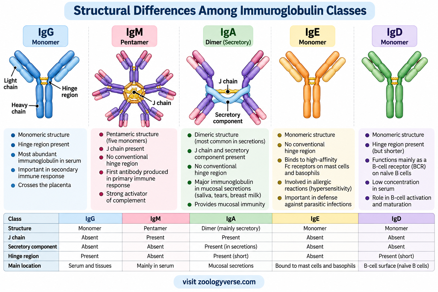
Although all five major immunoglobulin classes share the basic antibody design, they differ in their heavy-chain constant regions, number of domains, flexibility, molecular arrangement and distribution in the body. These structural differences influence where each class is found and how it contributes to immune defense.
The five major classes are IgG, IgM, IgA, IgE and IgD. Their classification depends on the type of heavy chain present: gamma (γ) in IgG, mu (μ) in IgM, alpha (α) in IgA, epsilon (ε) in IgE and delta (δ) in IgD.
IgG: Monomeric Structure
IgG is a monomer consisting of two heavy chains and two light chains. Each heavy chain contains one variable domain and three constant domains, while each light chain contains one variable and one constant domain.
The two Fab arms provide two antigen-binding sites, and the Fc region is formed mainly by the paired CH2 and CH3 domains. IgG contains a hinge region that allows movement of the Fab arms, although hinge properties differ among its subclasses.
IgG is the predominant immunoglobulin class in serum and has several subclasses in humans: IgG1, IgG2, IgG3 and IgG4. These subclasses differ in properties such as hinge structure, flexibility, complement activation and interaction with Fc receptors.
Its monomeric structure supports a range of functions, including neutralization, opsonization and, for suitable subclasses and immune complexes, complement activation. IgG can also cross the placenta through FcRn-mediated transport, providing passive immune protection to the developing fetus.
IgM: Pentameric Secreted Form
IgM has a heavy chain containing four constant domains (CH1–CH4) and lacks a conventional hinge region. This arrangement gives it a different structural organization from IgG.
Membrane-bound IgM is present on the surface of many mature naive B cells as part of the B-cell receptor. In this form, it is a membrane-associated immunoglobulin rather than a secreted pentamer.
Secreted IgM commonly forms a pentamer composed of five antibody units linked through disulfide bonds and associated with a joining (J) chain. This arrangement can provide up to ten antigen-binding sites, although not all sites are necessarily accessible or occupied at the same time.
Its multivalent structure makes IgM effective at agglutination and can support strong complement activation when the antibody is appropriately bound to a target. IgM is often produced early in a primary immune response.
IgA: Monomeric and Dimeric Forms
IgA contains alpha heavy chains and has three constant heavy-chain domains together with a hinge region. In serum, IgA is commonly found as a monomer. At mucosal surfaces, secreted IgA is often present as a dimer.
The dimeric form contains two IgA units associated through a J chain. During transport across epithelial cells, it acquires a secretory component derived from the polymeric immunoglobulin receptor. This component helps protect secretory IgA from enzymatic degradation.
The structure of secretory IgA supports immune defense at mucosal surfaces, including the respiratory and gastrointestinal tracts. It can neutralize pathogens and toxins and help prevent microorganisms from attaching to epithelial cells.
In humans, IgA has two subclasses, IgA1 and IgA2, which differ in structural features including their hinge regions.
IgE: Four Constant Domains and No Conventional Hinge
IgE contains epsilon heavy chains with four constant domains. Like IgM, it lacks a conventional hinge region and has an additional constant domain in its heavy chain.
IgE binds with high affinity to FcεRI receptors on mast cells and basophils. When an antigen cross-links receptor-bound IgE molecules, these cells can release inflammatory mediators. This mechanism is important in immediate allergic reactions and also contributes to defense against certain parasitic worms.
The structure of IgE allows it to interact with its receptors in a way that supports long-lasting receptor association. However, the amount of IgE circulating freely in serum is normally much lower than that of IgG.
IgD: Monomeric Structure with an Extended Hinge
IgD contains delta heavy chains, each with three constant domains and a relatively extended hinge region. It is mainly found on the surface of mature naive B cells, where it functions as part of the B-cell receptor alongside IgM.
Its hinge contributes flexibility, and its constant region has structural features distinct from those of IgG and IgA. Although IgD is present at low concentrations in serum, its membrane-bound form participates in B-cell antigen recognition and activation.
Antibody Structure: Labeled Diagram
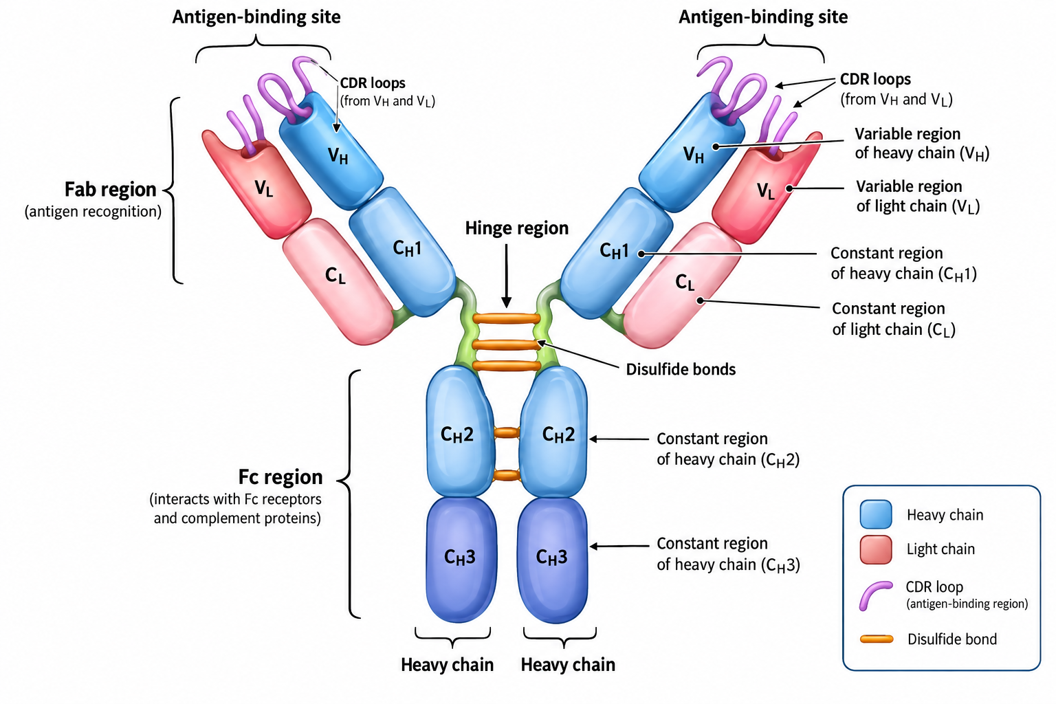
A labeled antibody diagram helps connect the names of its structural parts with their positions in the molecule. The conventional textbook model is a Y-shaped IgG antibody, which clearly illustrates the relationship between the antigen-binding arms, the hinge and the Fc region.
When studying or labeling an antibody diagram, remember that the drawing represents a simplified structural model. Other immunoglobulin classes, particularly secreted IgM and secretory IgA, have different overall arrangements.
Essential Labels in an Antibody Diagram
1. Heavy chains: The two longer polypeptide chains form the main framework of the molecule. They contribute to both Fab arms and the Fc region.
2. Light chains: The two shorter polypeptide chains associate with the heavy chains in the Fab arms. Each light chain contains a variable domain and a constant domain.
3. Variable regions (VH and VL): These are located near the tips of the Y-shaped molecule. The variable domains of one heavy and one light chain combine to form each antigen-binding site.
4. Constant regions: These domains support the antibody’s structural organization. The constant heavy-chain domains also help determine class-specific properties and effector interactions.
5. Antigen-binding sites: These are located at the ends of the two Fab arms. They recognize epitopes through complementary molecular interactions.
6. Fab region: Each arm contains the variable domains and the first constant domains of one heavy chain and one light chain. Fab is responsible for antigen recognition.
7. Fc region: The stem is formed mainly by the paired constant domains of the heavy chains. In IgG, it consists primarily of CH2 and CH3 domains and participates in interactions with Fc receptors and complement proteins.
8. Hinge region: In IgG, the hinge lies between CH1 and CH2 and allows movement of the Fab arms. It also contains inter-heavy-chain disulfide bonds in an arrangement that varies by subclass.
9. Disulfide bonds: These covalent bonds stabilize individual domains and connect the heavy and light chains. Their exact arrangement differs among immunoglobulin classes and subclasses.
10. CDR loops: These variable-region loops form much of the antigen-binding surface. They are not separate chains but parts of the folded VH and VL domains.
How to Interpret the Diagram
Start at the tips of the two arms and identify the antigen-binding sites. Follow each arm toward the centre to locate the variable and constant domains of the heavy and light chains. The arms meet the lower heavy-chain constant regions around the hinge area, and the paired heavy chains extend down to form the Fc stem.
The diagram should make the distinction between antigen recognition and effector activity clear: Fab binds the antigen, while Fc interacts with components of the immune system.
For exam purposes, label the heavy chains, light chains, variable and constant regions, Fab, Fc, hinge, antigen-binding sites and disulfide bonds. If the question asks specifically for IgG, show the conventional Y-shaped monomer rather than the pentameric structure of secreted IgM.
Relationship Between Antibody Structure and Function
The structure of an antibody determines how it recognizes antigens, maintains molecular stability and participates in immune defense. Its different regions work together, allowing one molecule to combine specific antigen recognition with interactions that help eliminate or control the target.
The relationship between structure and function can be understood through the following principles.
Structural Features and Their Biological Roles
| Structural feature | Relationship with function |
|---|---|
| Variable domains (VH and VL) | Form the antigen-recognition surface and determine binding specificity. |
| CDR loops | Provide much of the molecular diversity needed to recognize different epitopes. |
| Constant domains | Maintain the antibody framework and contribute to class-specific properties. |
| Disulfide bonds | Stabilize immunoglobulin domains and connect polypeptide chains. |
| Hinge region, when present | Provides flexibility that helps the antigen-binding arms accommodate different epitope arrangements. |
| Fc region | Enables interactions with Fc receptors and, under suitable conditions, complement proteins. |
| Multimeric organization | In secreted IgM and IgA, supports multivalent binding and specialized immune protection. |
| Fc glycosylation | Influences Fc conformation and interactions with immune-system components. |
How These Features Work Together
Antibody activity begins with the recognition of an appropriate epitope by the variable regions. The strength and specificity of this interaction depend on the shape and chemical properties of the antigen-binding surface. Structural stability allows these regions to maintain the arrangement required for effective binding.
Once an antibody attaches to its target, its remaining structural features influence the outcome. Depending on the immunoglobulin class, subclass and biological context, the bound antibody may block a pathogen or toxin, promote phagocytosis, recruit complement proteins or help immune cells destroy a target.
Differences between immunoglobulin classes further demonstrate this relationship. IgG is suited to diverse functions in blood and tissues, secretory IgA is adapted for mucosal protection, and the multivalent structure of secreted IgM supports agglutination and efficient complement activation.
Antibody structure provides the foundation for immune specificity and determines how that recognition is translated into biological activity. The coordinated arrangement of variable regions, constant domains, disulfide bonds and class-specific structural features makes antibodies versatile molecules of adaptive immunity.
Frequently Asked Questions (FAQs)
What is the basic structure of an antibody?
Why is an antibody called a Y-shaped molecule?
How many antigen-binding sites does an IgG antibody have?
What is the role of disulfide bonds in antibodies?
Do all antibodies have a hinge region?
Which part of an antibody determines its class?
What is the difference between an antibody and a B-cell receptor?
Why is the antibody structure important in immunity?
What is the difference between a monomeric and a multimeric antibody?
What is the function of the Fc region?
Key Takeaways
- A typical IgG antibody is a Y-shaped glycoprotein composed of two heavy chains and two light chains.
- The four polypeptide chains are connected and stabilized by disulfide bonds and noncovalent interactions.
- Each heavy chain contains one variable domain and three constant domains in IgG.
- Each light chain contains one variable domain and one constant domain.
- The two antigen-binding sites are formed by the association of the VH and VL domains.
- Complementarity-determining regions (CDRs) contribute substantially to antigen-binding specificity.
- The Fab region is responsible for antigen recognition, whereas the Fc region mediates many antibody effector interactions.
- Intrachain disulfide bonds stabilize immunoglobulin domains, while interchain bonds connect separate polypeptide chains.
- The hinge region provides flexibility in IgG, IgA and IgD; IgM and IgE lack a conventional hinge.
- The constant region of the heavy chain determines the immunoglobulin class.
- The five major immunoglobulin classes are IgG, IgM, IgA, IgE and IgD.
- Secreted IgM is usually pentameric, while secretory IgA is commonly dimeric.
- Fc glycosylation influences the structure and biological interactions of IgG.
- Antibody function depends on the coordinated activity of antigen-binding regions, constant domains and class-specific structural features.
Conclusion
Antibody structure is closely linked to the ability of the immune system to recognize antigens and respond to potential threats. A typical IgG molecule contains two heavy chains and two light chains arranged into antigen-binding arms and an Fc region, with disulfide bonds helping maintain its structural stability.
The variable domains provide antigen specificity, while the constant domains contribute to molecular organization and class-specific immune functions. Differences in hinge regions, heavy-chain domains and overall molecular arrangement distinguish IgG, IgM, IgA, IgE and IgD.
Understanding these structural features provides a foundation for studying antibody-mediated immunity, diagnostic techniques, vaccine responses and therapeutic antibodies. It also demonstrates how a common molecular framework can support a wide range of specialized immune functions.
References
- Janeway, C. A. Jr., Travers, P., Walport, M., and Shlomchik, M. J. Immunobiology: The Immune System in Health and Disease. 5th edition. Garland Science, 2001. Chapter 3: Antigen Recognition by B-cell and T-cell Receptors. NCBI Bookshelf – The Structure of a Typical Antibody Molecule.
- Alberts, B., Johnson, A., Lewis, J., et al. Molecular Biology of the Cell. 4th edition. Garland Science, 2002. Chapter 24: B Cells and Antibodies. NCBI Bookshelf – B Cells and Antibodies.
- Merck Manual Professional Edition. Molecular Components of the Immune System. Antibodies and Immunoglobulin Structure.
- StatPearls. Immunoglobulin. NCBI Bookshelf. Basic Immunoglobulin Structure and Function.
- Janeway, C. A. Jr., Travers, P., Walport, M., et al. Immunobiology: The Immune System in Health and Disease. NCBI Bookshelf – Antigen Recognition by B-cell and T-cell Receptors.
Discover more from Zoologyverse
Subscribe to get the latest posts sent to your email.