
Dissection of Pigeon : External Features
In order to study external morphology, take freshly chloroformed pigeon. Note that entire body, except the beak and feet, is covered by feathers. Body is regionated into head, neck, trunk and tail. The size and colour of the body vary in different species and varieties.
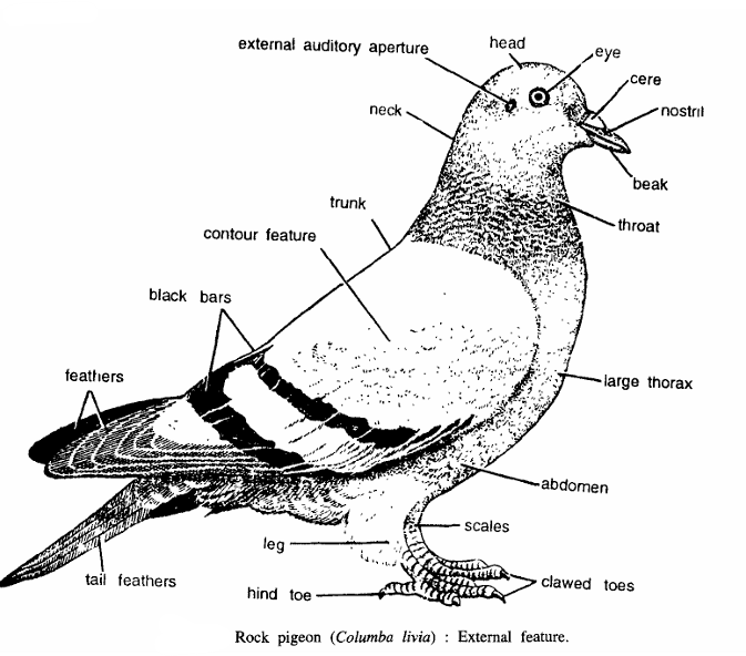
- Head :- It contains several structures :
- Beak or bill :- It is strong, horny, straight and pointed. At the base of the beak is a naked swollen portion of sensitive skin called cere.
- External nares or nostrils :- These are obliquely situated in the cere.
- Eyes and eyelids :- Each eye is large and rounded.
- External auditory aperture :- It is posterior to each eye and is covered by backwardly directed feathers, the auriculars.
- Neck :- It is long, flexible and S-shaped. Ventral part of neck is called as throat.
- Trunk :- It constitutes major part of the body. It is divided into a large thorax and a small abdomen. The thorax is strengthened ventrally by the large sternum, from which projects carina or keel. The keel is seen and felt as a prominent ridge. Trunk contains 2 pairs of limbs.
- Forelimbs :- These are modified into wings. Each wing is divided into the upper arm, forearm and hand.
- Hind limbs:- They support the body and are modified for bipedal locomotion. The hind limbs contain horny scales and each digit has a horny claw.
- Tail :- The region behind cloaca is called tail. The dorsal surface of the tail contains a uropygial gland secreting oil. Squeeze this gland and note oil oozing from it. The oil is used to dress or preen its feathers.
- Feathers :- Feathers cover more or less the entire body. They are of three kinds :
- Contour feathers. They cover the surface of the body consisting of wing feathers and contour feathers. The strongest contours are the quill feathers. The quill feathers of wings are called remiges and of tail as rectries. The remiges attached to bones of hand are called primaries and to ulna as secondaries. A small bunch of feathers is attached to the first pre-axial digit called alaspuria or bastard wing.
- Hair-feathers or jiloplumes. These are very thin, hair-like and degenerate feathers, situated at the base of the contour feathers.
- Down feathers. They are found below contour feathers and do not contain hooks on the barbules.
Dissection of Pigeon : General Anatomy
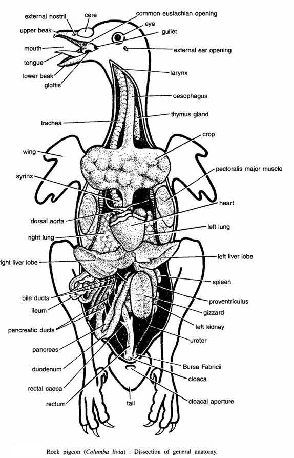
Procedure :- Kill the pigeon by chloroform. Remove the feathers by plucking them. After complete defeathering, lay the pigeon on its back on the dissecting dish. Cut longitudinally the abdominal wall from the cloaca. By strong scissors make a cut through the sides of sternum along its attachment to the ribs and lift the sternum with the fingers, so as not to injure the internal organs. Disarticulate the coracoids from the sternum and also cut the furcula at the two clavicles. The sternum can be detached to expose fully the underlying organs. Draw diagrams of organs seen in situ.
- Bucco-pharyngeal cavity :- It consists of :
- Horny beak :- Consisting of upper and lower beaks enclosing mouth.
- Internal nares :- These are slit-like openings, lying close together in the roof.
- Eustachian opening :- It is a single aperture found just behind the internal nares.
- Tongue :- It is triangular and pointed.
- Glottis :- It lies posterior to the tongue.
- Digestive system.
- Oesophagus :- It is large, wide and elongated and it opens into stomach. It extends through neck dorsal to trachea and forms a crop in the middle.
- Stomach. It comprises of a small digestive proventriculus and a large mechanical gizzard which is highly muscular.
- Intestine. Stomach leads into the intestine, which is differentiated into the duodenum, ileum and rectum. Rectum opens to outside by cloacal aperture.
- Associated glands and other structures. These comprise of liver, pancreas and spleen dorsal aorta, bile ducts, pancreatic ducts, Bursa Fabricii, ureter, left kidney, gizzard, proventriculus, spleen, left lung, heart pectoralis major muscles and thymus gland.
- Respiratory system. It consists of :
- Upper larynx. It opens into the pharynx by glottis.
- Trachea. It is situated ventral to the oesophagus and is supported by partially ossified rings. The trachea leads into right and left lungs and air sacs by bronchii. .
- Air sacs. Insert a blow pipe in trachea and inflate the air sacs. Tie a thread in trachea and examine the following in air sacs :
- Cervical air sacs. Two, found at the base of the neck.
- Interclavicular air sac. It is a single air sac, found between the two clavicles.
- Anterior thoracic air sacs. Two in number, covering the ventral surface of the anterior parts of the lungs.
- Posterior thoracic air sacs. Two in number, lying on the posterior parts of the lungs.
- Abdominal air sacs. Two in number. These are the largest and found dorsal to intestine.
Dissection of Pigeon : Flight Muscles
Procedure. In a defeathered pigeon, fixed in a dissecting dish, make a longitudinal incision in the skin from the cloacal aperture up to the lower beak. From the ends of the longitudinal incision cut the skin along the wings and pin up the flaps of skin or remove it by cutting. Also cut through the origin of pectoralis major by the side of keel, the posterior edge of sternum and the clavicle and from the underlying muscles. Study the following flight muscles :
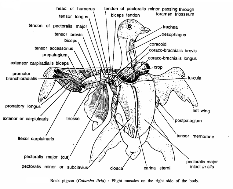
- Pectoralis major :- It originates from whole of the keel from the posterior part of the sternum and clavicle. Its flat tendon is inserted on the deltoid ridge of the humerus. Pectoralis major lowers the wing during flight. Hold the pigeon in your hand, pull the pectoralis major muscle and see how the wing is lowered.
- Pectoralis minor :- It overlaps the pectoralis from the dorsal part of the keel of sternum and the inner part of the ventral surface of the sternum. The tendon, formed by its fibres, passes through the foramen triosseum and is inserted on the dorsal surface of the humerus. Pectoralis minor raises the wings.
- Coraco-brachialis longus and coraco-brachialis brevis :- They originate from the coracoid and scapula and are inserted on the head of the humerous, to rotate the wings in glenoid cavity.
- Tensor longus, tensor brevis and tensor accessorius :- These hold the pre-patagium tensely stretched during flight.
- Tensor posterioris alae :- It keeps the post-patagium tensed. Other muscles associated with the flight are flexor carpiulnaris, extensor carpiulnaris, pronator longus, pronator brachioradialis, extensor carpiradialis biceps.
- Other structures seen are biceps, head of humerus, tendon of pectoralis minor passing through foramen triosseum, trachea, oesophagus, coracoid, crop, fercula, left wing, post, patagium, carina sterni and cloaca.
Dissection of Pigeon : Venous System
Procedure. Remove the pericardium and also cut away the fat and connective tissues at the base of the heart to expose the heart and blood vessels. Heart. It is four-chambered, consisting of two auricles and two ventricles. Veins. The blood from all parts of the body is collected by the following veins
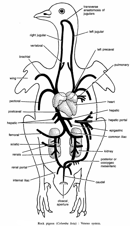
- A pair of pulmonary veins :- Two in number. They bring oxygenated blood from the lungs into the left auricle.A pair of pre-caval veins. Each pre-caval is formed by :
- Jugular vein :- Collects blood from the head. The right and left jugular veins unite in the neck by the inter-jugular transverse anastomosis. Both right and left jugular veins in middle give rise to right and left vertebral respectively which collect blood from vertebrae.
- Subclavian vein :-It receives a branch, the brachial, from the wing and a very large pectoral vein from the flight muscles.
- The post-caval vein :- It collects blood from the posterior part of the body and comprises of the following veins :
- Caudal vein :- Collects blood from tail.
- Renal portal or hypogastric vein :- The caudal vein bifurcates into two renal portal veins, which pass over the kidneys. Each renal portal vein receives :
- Internal iliac :- Collects blood from the pelvic region.
- Renal veins :- From the kidney.
- Femoral vein :- From outer side of leg.
- Sciatic vein :- From inner side of leg.
- Common iliac veins :- The above veins and renal portal vein on each side unite to form a common iliac vein :- The common iliac veins of two sides unite together to form the post-caval vein, which receives hepatic vein from the liver and enters directly into the right auricle.
- Hepatic portal vein. It collects blood from the intestine and it also receives a branch from the caudal vein called coccygeo-mesenteric vein. The hepatic vein also receives an epigastric vein from the peritoneum.
Dissection of Pigeon : Arterial System
The following arteries supply blood to various parts of body. Conus arteriosus and ventral aorta divide to form pulmonary aorta and right systemic arch.
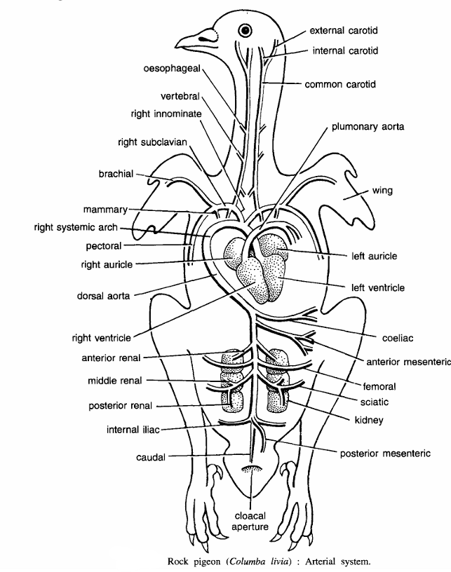
- Pulmonary arteries :- The pulmonary aorta divides into two pulmonary arteries, which carry deoxygenated blood to lungs, one on each side.
- Right systemic arch :- In birds only right systemic arch persists in the adult. It originates from the left ventricle and gives several arteries :
- A pair of innominate arteries. On each side, right systemic arch gives an innominate artery which gives two branches :
- Carotid artery :- It extends upwards and divides into external carotid and internal carotid to supply to the head.
- Subclavian artery :- It divides into brachial artery, which supplies to the wing, and pectoral artery, which supplies to the pectoral muscles of the flight.
- Dorsal aorta :- The right systemic arch bends over the heart and continues to posterior end as dorsal aorta, giving the following arteries :-
- Coeliac artery. It supplies to stomach and liver.
- Anterior mesenteric artery. It supplies to small intestine.
- Renal arteries. They supply to kidneys.
- A pair of femoral arteries. They supply to outer side of legs.
- A pair of sciatic arteries. They supply to inner side of legs.
- Iliac arteries. They supply to the hip region.
- Posterior mesenteric artery. It supplies to the large intestine.
- Caudal artery. It supplies to the tail.
- A pair of innominate arteries. On each side, right systemic arch gives an innominate artery which gives two branches :
Image Refernces :- Practical zoology Vertebrate , Dissection of Frog

I visited several blogs and i want say i just love your content. Thanks for sharing such informative articles.