
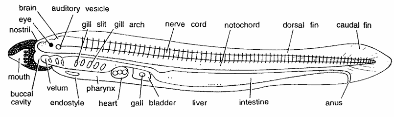
Lamprey : Ammocoete Larva Whole Mount
Comments :
- It is one of the stages in the development of lamprey.
- Egg develops into ammocoete larva, which is a tiny transparent creature in the beginning and later on becomes opaque and 170 mm long.
- It lies buried in mud.
- General body form is eel-like.
- Body divisible into head, trunk and tail.
- Mouth or buccal cavity is surrounded by upper and lower lips, having a number of buccal tentacles or oral cirri.
- Velum is found at the posterior end of the buccal cavity followed by the pharynx.
- Pharynx has 7 pairs of gill-slits. Gill arches and gill lamellae lie in the wall of these pouches.
- Ventrally pharynx contains double strand of mucus-secreting cells called as endostyle which forms thyroid gland of the adult. Head contains median nostril, eyes on sides and an auditory organ and brain.
- Larva contains median fin which forms continuous dorsal, caudal and ventral fins.
- Nerve cord with anterior brain divisions extends antero-posterioriy along the notochord. Notochord extends along the entire length of the body.
- Myotomes are segmentally arranged. Heart lies ventrally posterior to the pharynx and has 3 chambers.
- Ammocoete larva Digestive system after pharynx consists of oesophagus, wider intestine, anal opening, liver and gall bladder.
- Ammocoete larva shows intermediary characters between Cephalochordata and Cyclostomata
Identification : Since the above larva contains seven pairs of gill-slits and all above characters, hence it is Ammocoete larva.
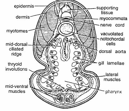
Lamprey : Ammocoete Larva : T.S. Passing Through Branchial Region
Comments :
- T.S. passing through branchial region shows distinct gill lamellae along with nerve cord and notochord, etc.
- Body wall is composed of epidermis, dermis and muscle layer. The muscles comprise of thick myotomes, separated by myocommata.
- Nerve cord lies just between supporting tissue and notochord.
- Notochord is surrounded by the notochordal sheath and has vacuolated chordal cells. Below notochord is wide-lumened pharynx. It contains several gill lamellae supported by lateral muscles.
- On dorsal side of the pharynx is dorsal aorta and mid-dorsal ciliated ridge, while on ventral side is thyroid involution.
- Below thyroid involution bundles of mid-ventral muscles are seen.
- Pharynx also contains velum, ciliated bands and endostyle.
- Pharynx performs both nutritive and respiratory functions.
- Endostyle in larval condition secretes mucus but during metamorphosis it develops into thyroid gland, which contains iodine vesicles
Identification : Since the larva contains mid-dorsal ciliated ridge and all above characters, hence it is T.S. of Ammocoete larva passing through branchial region.
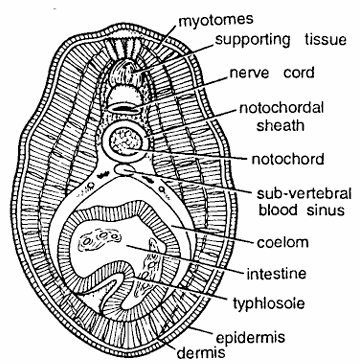
Lamprey : Ammocoete Larva : T.S. Passing Through Intestine
Comments :
- T.S. passing through the intestine shows usual parts of the body wall, nerve cord, notochord, muscles and intestine.
- Body wall is composed of epidermis, dermis and myotomes, which are especially developed on dorsal side.
- Supporting tissue and nerve cord lie close together.
- Vacuolated notochord is surrounded by notochordal sheath.
- Intestine is ventrally situated. It is lined by endodermal columnar cells.
- On ventral side intestinal epithelium is raised to form typhlosole, which increases absorptive surface.
- Sub-vertebral blood sinus is found below the notochord.
- Intestine is surrounded by coelomic cavity lined by coelomic epithelium
Identification : Since this section has typhlosole in intestine and all above characters, hence it is T.S. of Ammocoete larva passing through intestine.
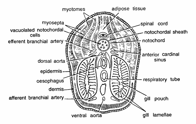
Petromyzon : T.S. Passing Through Branchial Region
Comments :
- T.S. passing through branchial region shows body-wall layers, prominent gill lamellae along with muscles, nerve cord, Notochord and other visceral structures.
- Body wall is composed of epidermis made up of stratified squamous epithelium, which is sometimes covered with polyhedral cells, dermis and muscle layer.
- Epidermal cells may be mucus-secreting cells, granular cells and club-shaped cells. Dermis is made up of a dense connective tissue of compact fibres.
- Muscle layer comprises of thick myotome bundles and myosepta or myocommata.
- Cut notochord is composed of vacuolated notochordal cells covered by notochordal sheath. Above notochord is nerve cord and adipose tissue, which forms fat column.
- Dorsal aorta is found below notochord followed dorsoventrally by oesophagus, respiratory tube and ventral aorta. Anterior cardinal sinus and efferent branchial artery are found on the sides of dorsal aorta in tandem position.
- Afferent branchial artery is found on each side of the ventral aorta.
- Two gills consisting of gill lamellae enclosing gill pouch occupy large space in the ventral half of the section.
Identification : Since this section contains large pharynx with gill pouches and all above structures, hence it is T.S. of Petromyzon passing through branchial region.
Petromyzon T.S. Passing Through Trunk Region
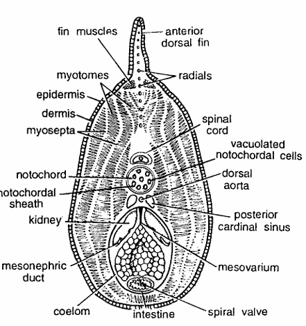
Comments :
- This section can be at once recognized by the presence of various visceral organs, containing body-wall layers, nerve cord, notochord, kidney, gonads and intestine, etc.
- Body wall comprises of polyhedral cells, stratified squamous epithelium, dermis and muscle layer.
- Epidermal cells consist of mucus secreting cells, granular cells and club-shaped cells.
- Dermis is made of dense connective tissue of compact fibres.
- Myotomes separated by myocommata are very distinct. Anterior dorsal fin is supported by cartilaginous radials arranged in a single series. Some fin muscles are also seen in the section.
- Vacuolated notochord is composed by notochordal tissue covered by notochordal sheath, which with its basement membrane forms elastic intema and a thin black elastic extema.
- Nerve cord is found above notochord. Also note posterior cardinal veins, dorsal aorta below notochord, and two kidneys with mesonephric duct.
- Gonad (ovary) is suspended from dorsal body wall by mesovarium. The intestine with spiral valve is also seen below ovary. Coelom is present.
Identification : Since this section contains visceral organs and all above features, hence it is T.S. of Petromyzon passing through trunk.
Petromyzon : T.S. Passing Through Tail Region
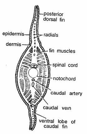
Comments :
- Section is smaller in outline.
- Body wall is composed of epidermis, dermis and muscles. (For details of body wall layers, see T.S. Petromyzon passing through branchial region).
- Dorsal and ventral lobes of the caudal fin are supported by cartilaginous radials.
- Spinal cord, notochord, caudal artery, and caudal vein are seen in the middle of the section.
- Myotomes and myosepta are distinct.
Identification : Series the section contains caudal fin and above features, hence it is T.S. passing through the tail region of Petromyzon.
