Amphibian slides of the frog are commonly used in zoology practicals to study the structural organization of a typical vertebrate. These slides often include transverse and longitudinal sections of skin, muscle, intestine, liver, kidney, ovary, testis, and blood vessels. Students examine frog slides to understand amphibian adaptations, organ systems, epithelial types, and basic vertebrate anatomy. Because frogs occupy an important evolutionary position between fishes and reptiles, these slides provide valuable insight into vertebrate physiology, tissue organization, and developmental biology.
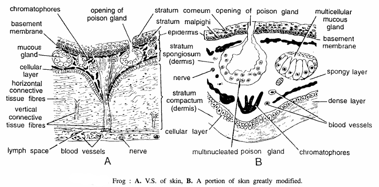
Amphibian Slides Frog V.S. of Skin
Comments Amphibian Slides Frog
- Under low magnification : (15 X eye-piece; 4 X objective).
- Vertical section passing through the skin reveals two layers, outer epidermis and inner dermis, distinctly seen under high magnification.
- Epidermis is thin and made up of startified squamous epithelium. Multilayered epidermis is differentiated into two layers :
- Outermost layer is stratum corneum and
- Inner layer called as stratum germinativum or stratum malpighi consisting of single layered closely placed nuclei. Beneath the epidermis is thin basement membrane and then dermis.
- Dermis constitutes major part of the skin and is largely made up of connective tissues. It is distinctly differentiated into two layers. Outer layer is made up of areolar spongy connective tissue and hence called as stratum spongiosum. It contains mucous glands, poison glands and chromatophores. Inner layer is made up of compact zigzag parallel fibres and hence called as stratum compactum. Dermis also contains few vertical fibers, blood vessels nerves and lymph space.
- A cellular layer separates stratum spongiosum and stratum compactum.
- Just beneath the stratum compactum is lymph layer and then body musculature. Connective tissue consists of horizontal connective tissue and vertical connective tissue.
- Under high magnification : (15 X eye-piece; 40 X objective) :- All the layers such as stratum corneum, stratum germinatum, stratum spongiosum and stratum compactum are clearly seen. Cells of stratum corneum are of varying sizes in the process of keratinization.
- Cells of stratum germinatum are rounded with a centrally placed nucleus and continuously mitoting. The poison gland has several nuclei (multinucleated) while mucous glands are multicelled structures. Both possess secretory granules. Most distinct structures seen are contracted and expanded chromatophores of ‘varying sizes. Vertical connective tissue fibres are also clearly seen.
Special features: Skin of frog performs various functions. It is protective, water observant, respiratory, mucous secreting, sensory, e:Acretory and swimming. Skin is an organ consisting of an ectodermal epithelium, the epidermis, and its supporting mesodermal connective tissue, the dermis. When the cells of stratum corneum die, they are shed off and this is called as moulting.
Identification: Since the section has thin epidermis and thick dermis and above features, hence it is V.S. of skin of frog.
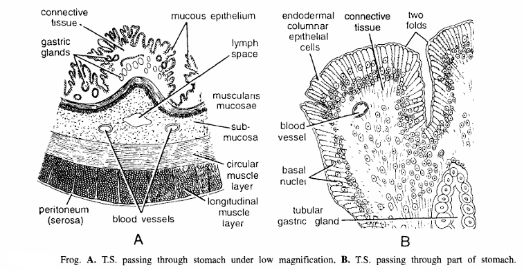
Amphibian Slides Frog T. S. Passing Through Stomach
Comments Amphibian Slides Frog
- Under low magnification: (10 X eye-piece; 4 X objective).
- Stomach is a broad tube, highly muscularised, having masticatory and digestive function. Histologically stomach is composed of serosa, muscle layer, sub-mucosa, muscularis mucosa and mucosa.
- Serosa forms the outermost thin layer. It is derived from visceral peritoneum and is composed of flat squamous cells, called as mesothelium.
- Muscle layer consists of outer longitudinal and inner circular muscle fibres.
- Outer longitudinal muscles run longitudinally and are made up of unstriped fibers. By the contraction of longitudinal muscles, stomach becomes shortened and the volume of lumen is widened.
- Inner circular muscles consist of circular fibers. By the contraction of these muscles, stomach increases in the size but the volume of the lumen is reduced.
- By alternate contractions and relaxations of longitudinal and circular fibers, the food is pushed backward and is also masticated.
- Sub-mucosa is made up of the loose areolar connective tissue. It serves to bind loosely muscularis mucosa with the muscular coat. It also contains blood vessels.
- Muscularis mucosa is a thin layer consisting of longitudinal and circular layers.
- Mucosa is the innermost layer thrown into folds. Mucosa consists of endodermal columnar epithelial cells, and connective tissue. Mucosa contains, oxyntic cells (HCI secreting), zymogen cells (pepsin secreting) and mucous cells (mucus secreting). The villi are of different sizes.
- Under high magnification: (10 X eye-piece; 40 X objective) :- Endodermal columnar epithelial cells devoid of goblet cells are clearly seen. Each cell is tall and having basal nucleus. Some of the cut tubular glands derived from mucosa are also seen. Substance of each villus contains several nuclei.
Special features (functions) : As soon as food reaches into the stomach, its muscular walls masticate the food; its gastric glands secrete digestive enzymes which hydrolyse the food. Pepsin breaks peptide bonds and converts proteins into derived proteins i.e., peptones and proteoses. HCI kills the bacteria or living food. The secretion of the gastric glands is under neurohormonal control.
Identification : Since the section has thick musculature and mucosal folds and above features, hence it is T.S. of stomach of frog.
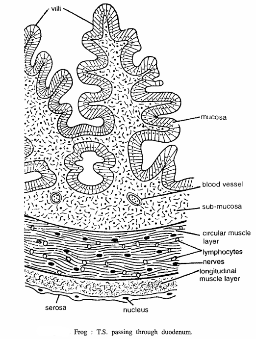
Amphibian Slides Frog T.S. Passing Through Duodenum
Comments
- Histologically duodenum resembles with ileum but its mucosa is peculiar. Section shows serosa, muscle layer, sub-mucosa, muscularis mucosa and mucosa.
- Serosa forms outer covering of duodenum. It is derived from the visceral peritoneum and is composed of flat squamous epithelial cells, called as mesothelium.
- Muscle layer is composed of an outer thinner longitudinal and inner thicker circular fibres.
- Longitudinal muscles are composed of unstriped fibres. By their contraction, the duodenal tube IS shortened but the volume of its lumen is widened.
- Circular muscles consist of circular fibres. By their contraction, the duodenal tube increases in size but the volume of its lumen decreases.
- Circular and longitudinal muscle layers contain nerves and lymphocytes.
- Sub-mucosa is well developed and is composed of loose connective tissue. In it the blood vessels and lacteals ramify before entering or after leaving the mucous membrane. It also contains nerves and blood vessels.
- Mucosa is thrown into irregular and branched villi.
Special features (functions) : Cholecystokinin and secretin, secreted in duodenum stimulate liver to secrete bile and pancreas to secrete pancreatic juice, respectively. In duodenum food is converted into amino acids and polypeptides, maltose and fatty acids and glycerol.
Identification : Since it has thin musculature, long and branched mucosal villi and above, hence it is T.S. of duodenum of frog.
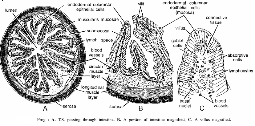
Amphibian Slides Frog T.S. Passing Through Intestine
Comments
- A. Under low magnification: (10 X eye-piece; 4 X objective).
- Intestine is modified for absorption and hence it is less muscularised. T.S. passing through it shows serosa, muscle layer, sub-mucosa, muscular is mucosa and mucosa.
- Serosa coat is complete except over part of the duodenum. Serosa originates from the visceral peritoneal layer and is composed of flat squamous epithelial cells, called as mesothelium.
- Outer longitudinal and inner circular muscle layers are distinct.
- Longitudinal layer consists of unstriped fibers. Size of the intestine is decreased and the volume of the intestinal lumen is increased by the contraction of the longitudinal layer.
- Circular muscle fibres form comparatively thick layer than longitudinal muscle layer. When they contract, size of the intestine is increased and the volume of the lumen is decreased.
- Between longitudinal and circular layers, a network of the lymphatic vessels and plexus myentericus, consisting of plexus of amyelinated nerve fibres, is found. However this layer is indistinct even in high magnification.
- Sub-mucosa is also well developed, made up of connective tissue and having nerves, rich lymph spaces and blood vessels.
- Blood vessels and lacteals ramify before or after leaving the mucous membrane. Nerve plexus is called as plexus of Meissner.
- Muscularis mucosa is not well developed.
- Mucosa is thrown into villi of different sizes. In a complete cut section the villi occupy most of the space of the lumen. Very small lumen is left. The branching of the mucosa increase~ the surface area of absorption.
- Under high magnification: (10 X eye-piece; 40 X objective).
- The mucosa is best seen under this magnification. It is thrown into many simple villi consisting of tall elongated endodermal columnar epithelial cells. Each cell has basal nucleus. Intermingled with epithelial cells are goblet cells and absorptive cells. Lymphocytes are also seen. Tubular glands are absent. The substance of villi contains several nuclei.
- Magnified villus shows endodermal columnar epithelial cells, goblet cells basal nuclei, blood vessels connective tissue and lymphocytes.
Special features (functions) : Goblet cells secrete mucous, while absorptive cells absorb food material. Intestinal enzymes are peptidases, maltase, lipase, invertase and lactase, which hydrolyse the food into amino acids sugar, fatty acids and glycerol, glucose and fructose and glucose and galactose, respectively.
Identification : Since it has goblet cells in mucosal folds, and all above features, hence it is T.S. of intestine of frog.
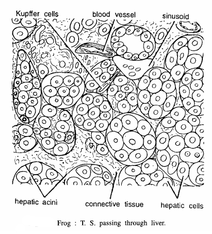
Amphibian Slides Frog T.S. Passing Through Liver
Comments
- Under low magnification: (15 X eye-piece; 4 X objective).
- Amphibian liver is dark chocolate coloured and large in proportion to the size of the body. The liver of frog contains three unequal lobes but connected with each other. T.S. passing through the liver shows the following histological details.
- It is a solid glandular organ, made up of rounded hepat.c lobules or acini in the form of branched columns, separated from one another by the connective tissue. Externally it is covered by serous coat.
- Each acini or hepatic lobule contains roughly 5 to 10 hepatic cells which are penetrated by fine network of connective tissue and sinusoid vessels called as hepatic capillaries.
- Portal vein, afferent blood vessel and hepatic artery enter its undersurface, where bile duct passes from the gland. Fine branches from above three form interlobular and intralobular vessels. Vessels are indistinct.
- Bile ducts and bile canaliculi are also seen.
- Each hepatic cell is rounded and nucleated.
- Glycogen formation occurs in the middle of the lobule, while peripheral area secretes bile.
- Large ducts drain bile capillaries and they unite to form hepatic duct.
- Bile secreted by liver is stored into large pear-shaped saccular structure, called as gall bladder.
- Under high magnification : (10 X eye-piece; 40 X objective).
- Hepatic acini and hepatocytes are very distinct. Each hepatocyte is rounded and contains one or two nuclei. Hepatocytes with two nuclei appear to be in mitotic stage. Cytoplasm is granular. Wall of the sinuses contains spindle-shaped Kupffer cells of phagocytic nature.
- Special features (functions) : Liver has the following functions :
- It secretes bile juice, consisting of bile salts, bile pigments, cholesterol and lecithin, which act as fat emulsifier.
- It stores glycogen, inorganic salts of iron and copper. Glycogenesis and glycogenolysis take place in liver,
- Liver produces fibrinogen and prothrombin, which are essential components of clotting of blood. It produces heparin, which prevents blood clotting,
- Liver changes ammonia into urea. Urea synthesis takes place by ornithine cycle. This process is also called as deamination.
- Liver also controls oxidation of sugar,
- It is excretory,
- In embryonic condition liver produces blood corpuscles,
- Various enzymes are synthesized in liver,
- Liver also stores and synthesizes vitamins,
- Liver is a very important organ for metabolism.
Identification: Since it has rounded hepatic lobules, and all above features hence it is T.S. of liver of frog.
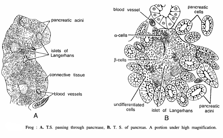
Amphibian Slides Frog T.S. Passing Through Pancreas
Comments
- Under low magnification: (7 X eye-piece; 4 X objective).
- Pancreas is noteworthy because in amphibians one or more pancreatic ducts open either directly into duodenum or indirectly through bile duct.
- Pancreas is a large and yellow-coloured compound tubulo-alveolar gland, situated between duodenum and stomach, and covered with coelomic epithelium.
- Histologically it is composed of several lobules or acini, connected by loose connective tissue, nerves and blood vessels. Pancreatic acini represent the secretory part of gland. Cut lobules may be rounded or tubular.
- Under low magnification the cellular organization is not very clear but the same could be clearly studied under high magnification.
- Large veins and arteris and several blood capillaries lie in the connective tissue.
- Centre of the acinus communicates with the nearest duct and it secretes digestive enzymes.
- Lightly coloured islets of Langerhans are scattered groups of cells surrounded by pancreatic acini.
- Central mass is peculiar and contains special cells, called as islets of Langerhans.
- Pancreas is very vascular and also innervated by parasympathetic nerves.
- Under high magnification : (15 X eye-piece; 40 X objective)
- Each lobule or acinus is made up of 13 to 15 cells. Wall of each acinus is made up of columnar or pyramidal cells. Each cell is differentiated into two zones- a basal zone containing rounded nucleus and some basophilic course granules and luminal zone with fine granules. In the scattered group of islets of Langerhans 3 kinds of cells are clearly seen –
- darkly stained alpha cells secreting glucagon,
- lightly stained rounded Beta cells secreting insulin and
- dot-shaped undifferentiated cells of unknown function. Cells of lobules may be squamous or cuboidal.
- Each lobule or acinus is made up of 13 to 15 cells. Wall of each acinus is made up of columnar or pyramidal cells. Each cell is differentiated into two zones- a basal zone containing rounded nucleus and some basophilic course granules and luminal zone with fine granules. In the scattered group of islets of Langerhans 3 kinds of cells are clearly seen –
Special features (functions) : Pancreas plays a dual role in the body, serving both as exocrine and endocrine glands. Exocrine secretion consists of digestive enzymes, such as amylopsin, trypsin and lipase. Endocrine secretion is that of islets of Langerhans which produce insulin and glucagon, secreted by alpha cells and beta cells, respectively. Insulin plays an important role in carbohydrate metabolism. It regulates blood sugar level. Its deficiency causes a disease, called as diabetes. Glucagon increases the blood sugar level. Its deficiency causes hypoglycemia.
Identification : Since the above section contains pancreatic lobules and islets of Langerhans and all above, hence this is T.S. of frog passing through pancreas.
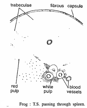
Amphibian Slides Frog : T.S. Passing Through Spleen
Comments
- Spleen is a small, rounded and dark brown structure, found above rectum.
- Spleen is covered with a fibrous and muscular capsule and externally covered by visceral peritoneum.
- Capsule sends bands or trabeculae, which ramify into the substance of the gland.
- Into the network of the trabeculae there is a soft pulpy mass, called as splenic pulp.
- Spleen T.S. shows presence of centrally located red pulp and white pulp.
Special features (functions) : (i) B-Iymphocytes produce antibodies, (ii) Spleen stores and synthesizes leucocytes (W.B.C.). Spleen is dilatable and contractile, (iii) It contains macrophages, which are responsible for the destruction of old erythrocytes. (iv) In embryonic condition it produces erythrocytes but after birth leucocytes are produced. blood vessel Frog: T.S. passing through spleen.
Identification : Since the above section contains red pulp, white pulp and trabeculae and all above features, hence it is T.S. of spleen of frog.
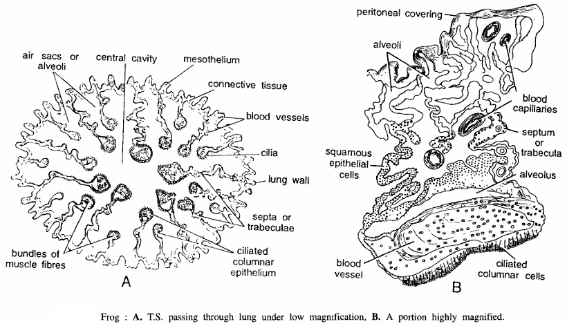
T.S. Passing Through Lung
Comments
- Under low magnification: (7 X eye-piece; 4 X objective).
- There is a pair of sac-like lungs, one on either side of heart. Lung of frog is not much elongated, but it is well developed having more alveolar spaces and rich blood supply.
- T.S. of lung shows peritoneal layer, lung wall, alveoli and a central cavity.
- Lung wall is composed of outer peritoneum made-up of squamous epithelial cells, connective tissue and unstriped muscle fibres. Inner wall is composed of ciliated epithelial cells, mucous glands and blood capillaries. Mucous glands are indistinct.
- Blood capllaries from network.
- Central cavity is partly divided into numerous chambers or alveoli, partitioned by trabeculae.
- Trabeculae partly contain flattened squamous epithelium and partly ciliated columnar epithelial cells. They are richly vascularised and contain several nuclei.
- Alveoli and trabeculae increase the respiratory surface.
- Under slightly high magnification : (7 X eye-piece; l0X objective).
- Squamous epithelial lining on trabeculae and expanded ciliated epithelial cells are very clear. Nuclei are concentrated on the sides of the trabeculae and the expanded cells. Other structures seen are mesotherium, connective tissue blood vessels, trabeculae, alveolis.
Special features (functions) : Lung is an important respiratory organ meant for external respiration, where oxygen from the atmosphere is taken to combine with haemoglobin forming oxyhaemoglobin. From the lungs oxyhaemoglobin is transported to cells for tissue or cellular respiration.
Identification : Since it has alveoli and air spaces and all above features, hence it is section of lung of frog.
T.S. Passing Through Spinal Cord

Comments
- A. Under low magnification: (7 X eye-piece; 4 X objective).
- Medula oblongata after emerging from foramen magnum, and continues posteriorly as spinal cord.
- T.S. of spinal cord shows that it is coated by outer durameter and inner piameter. Bioplasm of the nerve cord is divided by dorsal and ventral fissures and is differentiated into outer white matter and inner grey matter.
- White matter is devoid of nerve cells and represents lighter area.
- Grey matter contains nerve cells and a central canal, which is continued with the ventricles of brain. It is lined by a single epithelial ependyma. layer; called as Grey matter forms squarish area, but dorsal and ventral horns are not very much distinct.
- In mid-dorsal axis, dorsal and ventral fissures are seen.
- Under high magnification: (10 X eye piece; 40 X objective).
- Practically half of the outer peripheral portion of the spinal cord is occupied by the grey matter. Neurons are heavily concentrated. In mid dorsal axis, dorsal and ventral fissures are very clear.
Special features (function) : Spinal cord has the following functions : (i) It receives stimulus from dorsal and ventral roots, (ii) It transmits impulses to the brain, (iii) It causes reflex action.
Identification: Since it has white matter and grey matter and all above features, hence it is T.S. of spinal cord of frog.
T.S. Passing Through Kidney

Comments
- Under low magnification: (7 X eye-piece; 4 X objective).
- Kidney of adult frog is opisthonephros.
- There are two kidneys, each representing a compound tubular gland. T.S. passing through a kidney shows nephrostomes, uriniferous tubules and Bowman’s capsules.
- Each kidney is bean-shaped structure covered by visceral peritoneal layer.
- Interior of the kidney is filled with large number of uriniferous tubules cut in various planes.
- Nephrostomes are funnel-like structures communicating with the coelom and to the vein of kidney.
- Bowman’s capsules are cup-shaped structures, containing tuft of blood vessels called as glomerulus.
- Bowman’s capsule and glomerulus are collectively called as Malpighian body. These capsules are concentrated towards ventral region.
- Renal arteries and renal veins are cut at several places in section.
- Under high magnification : (10 X eye-piece; 40 X objective).
- Nephrostome or coelomic funnel is in the form of wide opening and it leads into collecting chamber and then into tubule. Other structures are Bowman’s capsule, glomerulus, uriniferous tubules, renal arteries and peritoneal covering.
- Portion of kidney with uriniferous tubules which are very much convoluted and made up of granular, ciliated and nucleated epithelial cells. Various uriniferous tubules cut in transverse and longitudinal plane are seen.
- Under high magnification shows double-walled Bowman’s capsule and glomerulus with wide afferent and narrow efferent network of capillaries forming glomerulus. The capsule has several nuclei of different sizes, blood vessels and squamous epithelial cells.
Function of kidney: It functions to stabilize blood stream by filtration extracting water, urea, uric acid, phosphates and sulphates, etc. Various diseases associated with kidney are glycosuria, albuminuria and nephritis, etc.
Identification : Since the section has glomerulus and Bowman’s capsule, hence it is T.S. of kidney of frog.
T.S. Passing Through Testis

Comments
- Under low magnification: (7 X eye-piece; 4 X objective).
- T.S. passing through testis shows that it is made up of peritoneal epithelium, tunica, albuginea, blood vessels, intertubular connective tissue and mesorchium.
- Testis are attached with kidney with mesorchium.
- Under high magnification : (15 X eye-piece; 40 X objective).
- T.S. of a seminiferous tubule shows that it is composed of a germinal epithelium which gives rise to spermatogonia or sperm mother cells.
- Other stages are spermatocytes, spermatids and sperms representing various stages of spermatogenesis are seen in the section.
- Section shows cut blood vessels and inter-tubular connective tissue.
- In section interstitial cells primary spermatocytes, secondary spermatocytes, spermatids and sperms are seen
- Interstitial cells present in the section secrete male hormone testosterone, which is responsible for developing secondary sexual characters.
- In high magnification :
- Various stages during spermatogenesis are clearly seen. The sperms mother cell or spermatogonia lie at the peripheral surface of seminiferous tubules. The primary spermatocytes are the largest cells having large nuclei.
- Secondary spermatocytes are smaller than primary having deeply stained nuclei. The spermatids are darkly stained still smaller rounded structure.
- Deep blue haematoxylin-stained spermatozoa with tails are present in large number. Intertubular connective tissue contains blood vessels and interstitial cells. Testis is coated with fibrous tunica albuginia and then by peritoneal epithelium.
Function : It produces sperms.
Identification: Since sections contains sperms and above features, hence it is T.S. of testis of frog.
Amphibian Slides Frog : T.S. Passing Through Ovary

Comments
- A. Under low magnification: (10 X eye-piece; 4 X objective).
- There are two ovaries attached to kidneys by mesovarium.
- Each ovary is composed of several hollow lobules containing developing ova in various stages of development, connective tissues, young follicles, blood vessel, primary oocytes germinal epithelium and theca.
- Under high magnification: (10 X eye-piece; 40 X objective).
- Each lobule is surrounded by theca extema, theca intema, germinal, epithelium, follicular cells and ova in various stages of development.
- An ovum under high magnification shows theca extema, theca intema, yolk granules, cytoplasm and nucleus.
Identification : Since A, B and C contains ova and above features, hence it is T.S. of ovary of frog.
Amphibian Slides Frog T.S. of Bone

Comments :
- Under low magnification : (10 X eye-piece; 4 X objective).
- T. S. bone shows outer periosteum, then outer zone of bone layer, inner zone of bone layer, bone marrow and central narrow cavity or canal.
- Under high magnification : (10 X eye-piece; 40 X objective).
- Outer most layer periosteum.
- Below periosteum is outer osteoblast layer.
- Beneath outer blast layer are lamellae osteocyte cells and inner osteoblast layer.
- Innermost in endosteum enclosing bone marrow.
Identification : Since the above section has osteocyte cells and above features hence it in T. S. of bone of frog.
Image References :- Practical zoology Vertebrate, Amphibian Slides
Discover more from Zoologyverse
Subscribe to get the latest posts sent to your email.