External Features
Dissection of Uromastix (Lizard) : Procedure :- Take a freshly-killed specimen and examine the external characters. Uromastix is commonly called as ‘Sanda’.
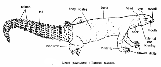
- Size :- It measures 20 to 30 cm in length.
- Shape :- Typically lizard like.
- Division :- Body is covered by epidermal scales and regionated into head, neck, trunk and tail.
- Head :- It is small with a short snout and contains the following structures :
- Nostrils :- The tip of the snout contains two large nostrils.
- Mouth :- It is a large opening, guarded by upper and lower jaws which contain cutting teeth, one in former and two in latter. Eyes. Eyes have two movable eyelids and a nictitating membrane.
- Openings of external ears :- Behind the eyes there are two sunken pits marking openings of external ears. Below each pit is a tympanic membrane, which shows beginning in reptiles of external auditory meatus.
- Neck :- It is short
- Trunk :- It is elongated forming major part of the body. It contains 2 pairs of limbs – fore and hind limbs. They are short and primitive and project laterally ending in 5 digits. Each digit contains a horny claw. The thigh region in males contains 12 to 18 pores of femoral glands on ventral surface. These glands produce a secretion, which hardens into temporary spines which are used for holding female during copulation.
- Tail :- The portion of the body behind cloaca is called tail. The tail is thick and contains about 27 rows of transversely arranged spiny scales. Hemipenis. The cloaca has a pair of copulatory hemipenis in male lizard only.
Arterial System of Uromastix (Lizard)
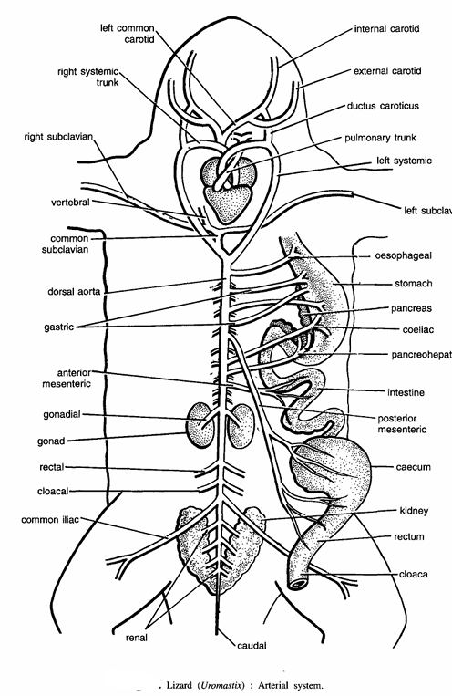
Dissection of Uromastix (Lizard) Procedure. Take a Uromastix freshly-killed with chloroform, wash it in water and lay it on its back in the dissecting dish. Pin the limbs of the lizard. Cover the dish with water. Make a longitudinal incision through the ventral body wall from the cloacal aperture up to the tip of the snout.
Cut the sternum and the pectoral girdle. Also make four cuts, two a little posterior to forelimbs and two opposite the hind limbs. Fix the flaps of the skin by pins. Cut very carefully the connecting membranes so as not to injure any organ and blood vessels. Cut the pericardium and trace the blood vessels, first veins and then arteries. The blood is distributed to all parts of body by the following arteries:
- Pulmonary arteries :- The pulmonary aorta originates from the right side of the ventricle and it divides into two pulmonary arteries, each going into a lung. They carry impure blood to lungs.
- Systemic arches :- The truncus arteriosus divides into right and left systemic arches. Right arch originates from the left side of the ventricle and left arch from the right side. Systemic arches give various arteries.
- Innominate artery :- It originates from the right systemic arch, which divides into the right carotid and left carotid arteries. Each carotid divides into an internal carotid and an external carotid.
- Ductus caroticus :- It joins the internal carotid artery with systemic arch of its side.
- Subclavian artery :- It originates from the right systemic arch and divides into right and left subclavian arteries which supply to arms. A vertebral branch from right subclavian supplies to vertebrae.
- Dorsal aorta. The right and left systemic arches unite together forming dorsal aorta, which proceeds backwards to supply by several arteries to viscera as follows :
- Oesophageal artery to oesophagus.
- Posterior mesenteric artery to caecum.
- Paired gastric arteries to stomach.
- Gonadial artery to gonad.
- Coeliac artery to stomach.
- Cloacal arteries to cloaca.
- Pancre-hepatic artery to pancreas and liver.
- Renal arteries to kidneys.
- Anterior mesenteric artery to ileum.
- Caudal artery to tail.
- Iliac arteries to legs.
Dissection of Uromastix (Lizard) : Venous System
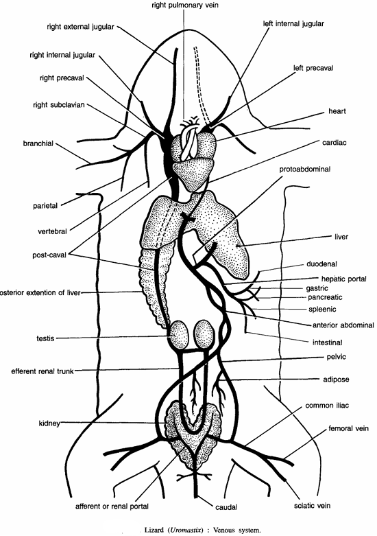
Dissection of Uromastix – The blood from the entire body is collected by the following veins :
- Pulmonary veins :- Collect oxygenated blood from lungs. They unite and open into the left auricle.
- Pre-caval veins :- Each pre-caval vein collects blood from the anterior end, by the following veins :
- External jugular :- Collects blood from head.
- Internal jugular :- Collects blood from head.
- Subclavian :- Collects blood from the arm. Note that there is no external jugular on the left side and left pre-caval contains only internal jugular and subclavian. The two pre-cavals enter the bilobed sinus venosus.
- Post-caval vein :- The blood from the posterior region is collected by post-caval vein, which pours blood into the sinus venous. The posterior veins are :
- Caudal vein. Collects blood from tail.
- Renal portal vein. The caudal vein divides into kidney to form renal portal veins.
- Femoral vein. Collects blood from outer side of leg.
- Sciatic vein. Collects blood from inner side of leg.
- Iliac vein. It is formed by the union of femoral and sciatic veins.
- Pelvic vein. On each side renal portal and iliac veins unite to form a pelvic vein. It also receives adipose vein from fat bodies.
- Anterior abdominal vein. The two pelvics imperfectly unite together to form anterior abdominal vein, which opens into the left lobe of the liver. Anterior abdominal continues towards heart as post abdominal and collects blood from heart by cardiac vein.
- Renal sinus vein. The capillaries in the kidney unite to form renal sinus vein, which extends forwards as efferent renal trunk.
- Post-caval vein. The two efferent renal trunks unite together below gonad and form the post-caval vein. The post-caval vein receives a wide hepatic vein from the liver, continues upward, and opens into the sinus venosus.
- Hepatic portal system :- It is formed by the following veins
- Gastric vein from stomach.
- Intestinal vein from intestine.
- Duodenal vein from duodenum.
- Splenic vein from spleen.
- Pancreatic vein from pancreas. These veins unite to form the hepatic portal vein, which joins with abdominal vein and enters the liver, where it breaks into capillaries forming hepatic portal system.
Image Refernces :- Practical zoology Vertebrate , Dissection of Frog