General Instructions for Dissecton of Scoliodon
For Dissecton of Scoliodon While dissecting the animal keep in mind the following points
- Follow carefully the instructions given by the teacher in your practical class.
- Study well about the internal structures of the animal to be dissected.
- Keep all the instruments in your dissecting box clean and sharp.
- Always keep with you a A text book of practical zoology vertebrate and also hand-drawn diagram of the dissection.
- Remember that all vertebrates are dissected from ventral side.
- Wash the animal before dissection to remove excess of formalin or other fixing or killing chemical.
- Keep a white sheet below the animal in dissecting dish. Fix the animal in dissecting dish properly. Insert the pins obliquely. While opening the animal never make deep incisions.
- Remove the body wall layers in such a manner that all the internal organs are fully exposed.
- Keep your dissection submerged in water.
- Remove unwanted tissue by cutting with scissors.
- Blackpaper your dissection. For example, keep small glazed black or blue paper below the nerves and nerve cord in dogfish.
- Flag label only when asked by the teacher. While doing it cut small flags, write the name of the organ or tissue and pin it with needle. Keep the flag near the blunt end of the needle and pin near the organ or tissue by pointed end of the needle obliquely.
- Always leave the dissection after cleaning the tissues in the dish.
- Display nicely the dissection.
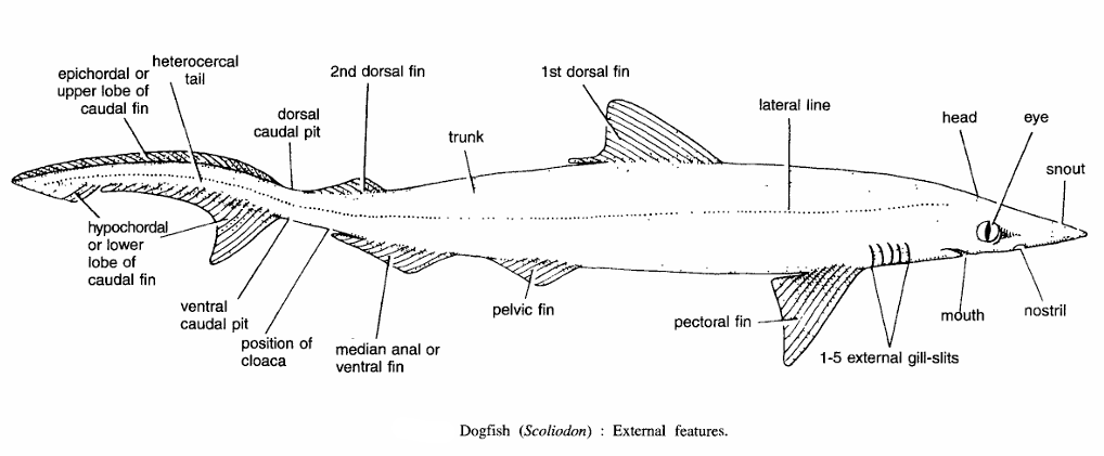
Dissecton of Scoliodon : External Features
Procedure.
Take a formalin-preserved or freshly-killed Scoliodon, wash in tap-water and examine the external features. Draw lateral view of the specimen. Observe and note the following features :
- Size :- Females are larger than males. Size in both specimens varies from 30 to 60 cm.
- Shape :- Spindle-shaped. Head is dorso-ventrally flattened, while rest of the body is laterally compressed.
- Colouration :- The back and sides of body are brownish in colour, while under-surface is yellowish white. Texture. The body surface is rough to touch due to backwardly-directed spines of the placoid scales.
- Divisions :- The entire body is regionated into head, trunk and tail.
- Head :- It is flattened and produced into snout in front of mouth. Note the following structures in head:
- Mouth :- It is crescentric in shape, found on the ventral surface and is guarded by the upper and lower jaws which contain backwardly-directed conical teeth.
- Nostrils :- These are obliquely-placed small apertures on either side of body, anterior to the mouth.
- A pair of eyes :- Each eye is large and situated on the lateral side of head. It contains upper eyelid, lower eyelid and a thin transparent nictitating membrane.
- Pores :- Several ampullary pores are present on the upper and lower surfaces of the head. Each pore opens into the lateral line canal.
- Gill-slits :- There are five gill slits on either side, a little behind the head.
- Trunk :- This region constitutes major part of the body starting from behind the gill-slits up to cloacal aperture. Trunk contains median unpaired fins and paired lateral fins.
- Median fins include :-
- First dorsal fin. It is found anterior to middle part of the body.
- Second dorsal fin. It is found a little behind the first dorsal fin and is a bit smaller than the first.
- Ventral fin. It is found on ventral side, little distance to the position of second dorsal fin.
- Paired fins are :
- A pair of pectoral fins. These are large fins found just behind the gill-slits and in horizontal plane.
- A pair of pelvic fins. These are very small and found on either side of cloacal aperture.
- Tail :-The portion of the body behind the cloaca is called tail. It propels the fish through the water by caudal fin. The caudal fin contains epicaudal and hypocaudal lobes. At the junction of caudal fin and tail is a caudal pit.
- Head :- It is flattened and produced into snout in front of mouth. Note the following structures in head:
A pair of claspers are found attached to the pelvic fins in males only. Sides of the fish contain faintly-marked lateral lines.
Dissecton of Scoliodon :- General Anatomy
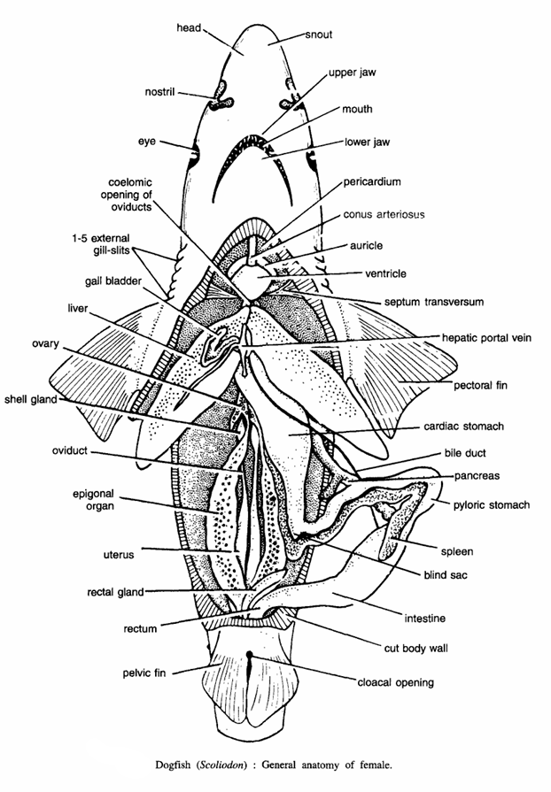
Procedure
Take a preserved specimen, wash in water and lay down on the dissecting board with the ventral surface upwards or facing towards you and fix the animal by pinning the pectoral fin. Make a mid-longitudinal incision in body wall from the cloacal aperture up to the pectoral girdle and also cut transversally at each end of the longitudinal incision. Reflect and pin down the cut flaps. Examine the following organs :
- Pharynx and oesophagus : Behind the pharynx is a short oesophagus which leads into the stomach.
- Stomach :- It is J-shaped and divided into the proximal cardiac stomach and distal pyloric stomach joined by a blind sac.
- Intestine :- Pyloric stomach opens into the wide intestine, which is internally folded into the scroll valve, which can be seen by cutting transversally or longitudinally.
- Rectum :- The intestine leads into cloaca. A large rectal or caecal gland opens on the dorsal surface of rectum.
- Liver :- It consists of two large and elongated lobes ventral to cardiac stomach. The two lobes are anteriorly jointed. Gall bladder is found on the dorsal lobe.
- Pancreas :- It is a bilobed compact gland, situated in the angle between two limbs of stomach.
- Spleen :- It is found in the coils of the pyloric stomach. Vascular organs. Structures seen are the pericardium, heart (auricle and ventricle), conus arteriosus, ventral aorta and septum transversum and hepatic portal vein.
- Male urinogenital organs :- These include testes, vasa deferentia, vesiculae seminales, sperm sacs, caecal gland, kidneys, ureters and cloaca.
- Female urinogenital organs :- The female specimen has ovaries, oviducts with oviducal funnels, epigonal organs, shell glands, uterus, caecal gland, kidneys, ureters and cloaca.
Dissecton of Scoliodon : Heart and Afferent Branchial Arteries

Procedure
For exposing heart and afferent branchial arteries, cut through the pectoral girdle and remove its middle portion. Take care not to injure the heart below. Remove the pericardium.
- Heart :- It is a dorso-ventrally bent muscular tube containing four chambers, namely sinus venosus, auricle, ventricle and conus arteriosus.
- Afferent branchial arteries :- The conus arteriosus is continued forward as ventral aorta which gives rise to afferent branchial arteries. The ventral aorta runs up to the posterior border of the pharynx. Distally it divides into two innominate arteries. Each innominate artery again divides into I afferent branchial artery and II afferent branchial artery. III, IV and V afferent branchial arteries originate directly from the ventral aorta at equal distances from each other. Five pairs of afferent branchial arteries supply to five pairs of gills.
Dissecton of Scoliodon : Efferent Branchial Arteries
Procedure
For efferent branchial arteries dissect the fish from the roof of the pharynx. There are 9 efferent branchial arteries on each side. The 1+11, 1I1+IV, V+VI and VII+VIII efferent branchial artery form four pairs of loops. The ninth efferent branchial artery joins with the VIII branchial artory. The four loops are connected together by longitudinal vessels. Each loop is continued as an epibranchial artery. Such as I epibranchial artery, II-epibranchial artery, III epibranchial artery and IV epibranchial artery. These four pairs of epibranchial arteries unite to form dorsal aorta. Also examine some main arteries supplying the head such as the afferent spiracular artery. Sub clavian artery, coeliacomesenteric artery and branches of dorsal aorta.
Dissecton of Scoliodon : Eye Muscles
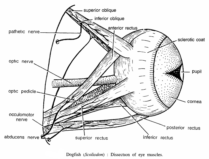
Procedure
Remove the eyelids and nictitating membrane to expose the eye-ball and its muscles. The eye muscles are inserted into the eye-ball in two groups. Note the following six eye muscles :
- Superior rectus :- Inserted on the dorsal surface of the eye-ball.
- Inferior rectus :- Inserted on the ventral surface of the eye-ball.
- Anterior rectus :- Inserted on the anterior surface of the eye-ball.
- Posterior rectus :- Inserted on the posterior surface of the: eye-ball.
- Superior oblique :- Inserted on the dorsal surface of the eye-ball.
- Inferior oblique :- Inserted on the ventral surface of eye-ball.
- Optic peduncle :- It is a cartilaginous stalk holding eye in the orbit.
Dissecton of Scoliodon : Internal Ear
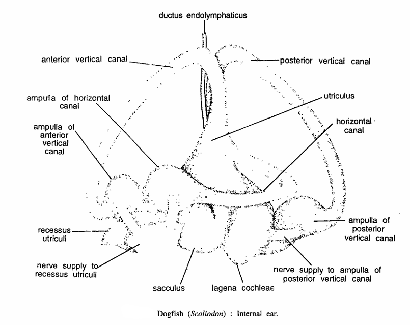
Procedure
The internal ear of membranous labyrinth lies in the auditor capsule just behind the orbit on either side. The auditory capsules are seen as bulgings on either side. Remove the skin over auditory capsules. Careful observation shows ridges of anterior vertical, horizontal and posterior vertical semicircular canals. The cartilage capsule can be gently broken by forceps. Take care not to injure the canals of membranous labyrinth. Locate the vertical canals and proceed. The internal ear consists of anterior vertical canal, posterior vertical canal, horizontal canal. ampullae of anterior vertical canal, horizontal canal and posterior vertical canal, lagena cochlea, recessus utriculi, utriculus and nerve supplies.
Dissecton of Scoliodon : Cranial Nerves
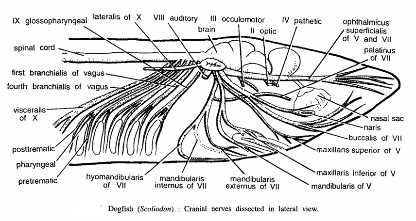
Procedure
Make a longitudinal incision in the middle of head from the gill cleft region up to the snout and also make transverse incisions on one side up to the lower edges. Remove the flap of skin. There are 10 pairs of cranial nerves emerging from the cranium as given in the following table :
| Name of the Nerve | Originates from | Nature | Innervation |
| (I). Olfactory | Olfactory lobe | Sensory | Nose Epithelium |
| (II). Optic | Optic Thalamus | Sensory | Retina |
| (III). Oculomotor | Ventral surface of mid brain | Motor | Eye Muscles (anterior rectus, superior and inferior rectii and inferior oblique) |
| (IV). Pathetic | Dorso-Iateral side of mid-brain | Motor | Superior oblique muscle of eye |
| (V). Trigeminal It has 5 branches | Side of medulla just below corpora restiformis | Mixed | |
(a) Opthalmicus profundus | Olfactory capsule and dorsal skin of snout | ||
| b) Opthalmicus superjicialis | Skin of snout | ||
| c) MaxilIaris superior | Skin of upper jaw | ||
| (d) Maxillaris inferior | Posterior part of upper lip | ||
| (e) Mandibularis | Muscles of lower jaw | ||
| (VI). Abducens | Ventral side of medulla | Motor | Posterior and external rectus muscles of eyes |
| VII. Facial It has the following branches : a).Opthalmicus superficialis b). Ramus buccalis c). Ramus hyomandibularis It has 3 branches : i). Mandibularis externus ii). Mandibularis internus iii). Hyoidean (d) Ramus palatinus | Cranium | ||
| VIII. Auditory | Side of Medulla close to V and VIII cranial nerves | Sensory | Internal ear |
| IX. Glossopharyngeal | Ventro-lateral side of medulla | ||
| X. Vagus | Side of medulla |
Well I really liked studying it. This subject provided by you is very effective for correct planning.