Fish slides of Scoliodon are widely used in zoology laboratories to study the internal and external anatomy of the spiny dogfish shark. These slides typically display structures such as the gills, fins, vertebrae, muscle blocks, and sensory organs. Students examine Scoliodon slides to understand cartilaginous fish morphology, respiratory adaptations, and evolutionary features that distinguish elasmobranchs from other vertebrates. Because Scoliodon represents a primitive yet well-organized fish, these slides help learners explore fundamental vertebrate anatomy and comparative morphology.
Transverse sections are prepared from specimens that have been preserved and hardened in formalin. With the help of a Butcher’s knife or sharp razor, cut the hardened specimen through eye region, olfactory region, branchial region, stomach and liver region, intestinal region, pelvic fin region and caudal region. Study various hand cut sections.
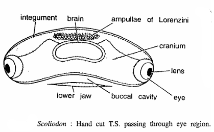
Fish Slides Scoliodon : Hand Cut T.S. Passing Through Eye Region
Comments Fish Slides Scoliodon
- This section can at once be recognized by prominent eyes on the sides.
- Entire section is covered by the integument, which contains several placoid scales and small denticles.
- Eyes are found on either side of the section. Each eye contains cornea and lens. Eye is innervated by thick optic nerve.
- Interior of the section is occupied by the cartilaginous cranium.
- In the middle of the section is brain.
- On the ventral side lower jaw is also seen.
- On the dorsal side several rounded sensory ampullae of Lorenzini are seen.
- Myotomes are absent.
Identification : Since this section contains cut eyes at both the ends, hence it is T.S. of Scoliodon passing through eye region.
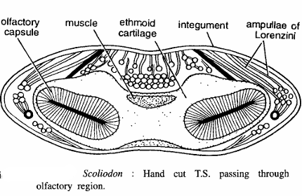
Hand Cut T.S. Passing Through Olfactory Region
Comments Fish Slides Scoliodon
- Section is somewhat oval in outline.
- It is covered by integument on all sides containing placoid scales.
- Middle of the section has muscles.
- On other side of the section olfactory capsules are seen.
Identification : Since this section contains cut olfactory capsules, hence it is T.S. of Scoliodon passing through olfactory region.
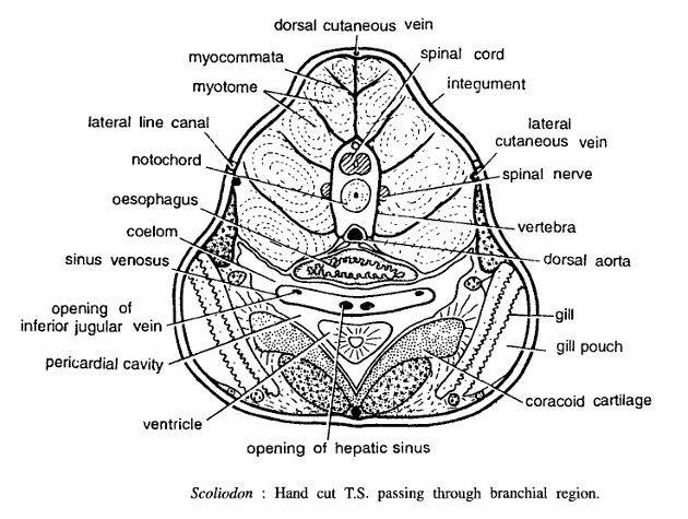
Scoliodon Hand Cut T.S. Passing Through Branchial Region
Comments Fish Slides Scoliodon
- T.S. passing through above region shows body wall layers, myotomes, spinal cord, notochord, oesophagus, heart and branchii or gills.
- Section is covered by pigmented integument from all sides composed of epidermis and dermis. Epidermis contains several dermal placoid scales.
- Dorsal half of the section has thick segmental muscles bundles or myotomes, separated by myocommata. Myotomes are concentrically arranged.
- In the middle of the muscle layer, cartilaginous vertebra containing spinal cord is found.
- Notochord is found just below the spinal cord and dorsal aorta is situated just beneath the vertebra.
- Oesophagus with thick walls and villi is found just below the dorsal aorta. Beneath oesophagus is pericardial cavity enclosing sinus venosus and the thick-walled ventricle. Below pericardium is the coracoid cartilage.
- Gills, with gill pouches, are found on the sides of pericardium and coracoid cartilage. Gills occupy entire side of ventral halves. Various cut blood vessels are also seen.
Identification : Since this section contains gills and all above structures, hence it is T.S. of Scoliodon passing through gill region.
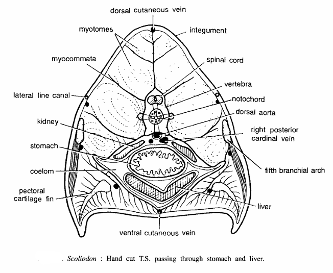
Scoliodon : Hand Cut T.S. Passing Through Stomach and Liver Region
Comments
- T.S. passing through above region shows body wall layers, muscle layer, spinal cord, stomach and liver. Section is roughly triangular.
- Body consists of integument and muscle layer. Integument is made of epidermis and dermis. Several dermal placoid scales are found in the highly pigmented integument.
- Myotomes are thick muscle bundles, arranged concentrically and separated by the myocommata. Practically entire dorsal half of the section contains segmental myotomes.
- In the central axis there is a median vertebra.
- Spinal cord with neurocoel is found in the vertebra and dorsal aorta just below the vertebra.
- Notochord is found just beneath the spinal cord.
- Ventral half of the section has a spacious coelomic cavity, lined by visceral and peritoneal layers and containing visceral organs.
- Thick-walled stomach with internal villi is found in the middle. Kidneys are found on the sides of stomach, while liver is found below the stomach.
- Cartilage of pectoral fin, cut portions of cutaneous veins, fifth branchial arch and lateral line canal are seen in the section.
Identification : Since this section contains cut stomach and all above structures, hence it is T.S. of Scoliodon passing through stomach and liver region.
Hand Cut T.S. Passing Through Intestinal Region
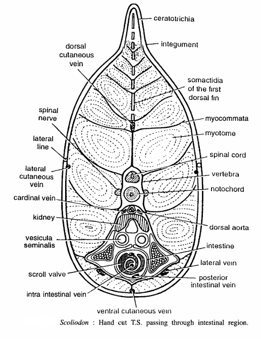
Comments
- T.S. passing through intestinal region shows body wall layers, spinal cord, notochord, kidneys, gonads, scroll valve in the intestine, etc. Section is pointed dorsally.
- Integument with dermal placoid scales covers the section from all sides. Body wall consists of epidermis, dermis and musculature.
- Myotomes arranged concentrically and myocommata occupy more than the dorsal half of the section on each side.
- Vertebra containing nerve cord or spinal cord and notochord is found in the median axis.
- Dorsal aorta is found just below the vertebra.
- Coelomic cavity is reduced. It accommodates viscera.
- Kidneys are found below dorsal aorta. Cut portions of the seminal vesicles and testes with spermatozoa are found below kidneys.
- Intestine is found below seminal vesicles. Internal fold forming anti-clockwise scroll valve is very distinct. Various cut blood vessels, spinal nerves and lateral lines are also seen in the section.
Identification : Since this section contains rolled intestine and above features, hence it is T.S. through the intestine of Scoliodon.
Fish Slides Scoliodon : Hand Cut T.S. Passing Through Base of Pelvic Fin of Male and Female
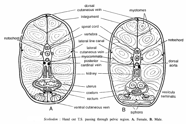
Comments
- T.S. passing through the pelvic region shows body wall layers, nerve cord, notochord, kidney, uterus in females and seminal vesicles in males, etc.
- Body wall is composed of integument and muscle layer. Integument covers the section from all sides. It consists of epidermis and dermis. Various dermal placoid scales are found in the skin.
- Muscle layer consists of concentrically arranged myotomes separated by myocommata and they occupy the entire section except some central area for viscera.
- Vertebra is found in the central axis. It encloses spinal cord and notochord in tandem position.
- Coelomic cavity is much reduced.
- Dorsal aorta is found just beneath the vertebra. Posterior cardinal vein, kidney and ureter are found below dorsal aorta, one after the other, towards posterior side.
- In female Scoliodon the two uterii fuse together forming a single uterus, which lies the ureters.
- In male Scoliodon two seminal vesicles lie below the ureters.
- Various cut portions of blood vessels, cartilage of pelvic fin and rectum are also seen in the section.
Identification : Since the above sections contain uterus in female and seminal vesicles in male, hence A and B are sections passing through the pelvic region of female and male Scoliodon, respectively.
Fish Slides Scoliodon Hand Cut T.S. Passing Through Caudal Region
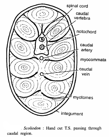
Comments
- T.S. passing through the caudal region shows body wall, caudal vertebra and muscle bundles. No other structure is found in this region.
- Body wall is composed of integument and muscle layer.
- Integument consists of epidermis and dermis. Dermal placoid scales present. The concentrically arranged myotomes separated by myocommata occupy almost entire space of the section.
- Vertebra is found in sub-equatorial mechanical axis. Spinal cord and notochord are enclosed in the vertebra.
- Caudal vein is found below the caudal artery and the latter is found below the vertebra.
Identification: Since the above section contains caudal vertebra and above features only hence it is T.S. of Scoliodon passing through the caudal region.
Image References :- Practical zoology Vertebrate