Mammal Slides or Rabbit slides are widely used in zoology and histology practicals to study the tissue structure and organ systems of mammals. These slides typically include transverse and longitudinal sections of skin, muscle, intestine, liver, kidney, lung, heart, ovary, and testis. Students examine rabbit slides to understand mammalian characteristics such as hair, endothermy, four-chambered heart, and highly developed organ systems. Because rabbits represent a typical small mammal with well-differentiated tissues, these slides help learners explore vertebrate anatomy, physiology, and comparative histology.
Rabbit mammal slides help students study mammalian tissues and organs, offering insight into vertebrate anatomy, physiology, and comparative histology.
Mammal Slides Rabbit : V.S. of Skin
- Mammalian skin is characterized by having various glands and hairs in abundance.
- T.S. through skin shows that it is an organ consisting of an ectodermal epithelium, the epidermis, and its supporting mesodermal connective tissue, the dermis.
- Epidermis constitutes the investing cellular membrane of the organisms. It is a stratified squamous epithelium which represents peculiar metabolic end product in the form of fibrous protein, keratin. The epidermis consists of the following layers :
- Stratum corneum : Its cells are a nucleate broad flat scales. These cells are the end product of the process of epidermal keratinization. Cells are bi-refringent containing disulphide linkages.
- Stratum lucidum : Next layer consists of flat cells without granules and having eleidin.
- Stratum granulosum : Transition from the stratum germinativum to the stratum corneum is marked by the stratum granulosum, a layer of flattened cells whose nuclei become pycnotic and whose cytoplasm contains refractory granules, the keratohyaline granules.
- Stratum malpighii : This is innermost layer, consisting of germinal layer. New cells are budded off from this layer mitotically and move upwards. Hairs are characteristic of mammalian skin only. Each hair is an elongated structure consisting of hair shaft, hair follicle, and hair pin.
- Hair shaft projects obliquely on the body surface. Erector muscles move the hair involuntarily.
- Dermis : Beneath epidermis is dermis. It is formed of areolar connective tissue. It originates from dermatome. It contains sebaceous glands and sweat glands.
- Below dermis is a sub-cutaneous reticular layer, consisting of penniculus adipose globules.
- Golgi mazzoni corpuscles, Pacinian corpuscles and endings of ruffini are also seen in the section.
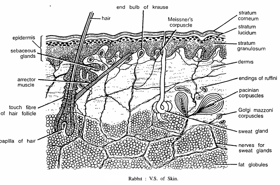
Special features : Skin serves various functions such as-protective, (ii) defensive, (iii) maintains shape, (iv) receives stimuli, (v) acts as insulators, (vi) prevents loss of water due to adipose tissue, (vii) excretory (sweat gland eliminates water and other waste products), (viii) milk producing (by mammary glands), etc.
Identification : Since section contains hair and above features, hence it is V.S. of skin of mammal.
Mammal Slides Rabbit T.S. Passing Through Cardiac Stomach
Comments
- A. Under low magnification: (7 X eye-piece; 4 X objective).
- Stomach is an important, sac-like structure, functioning digestive and masticatory. It is composed of serosa, muscle layer, sub-mucosa, muscularis mucosa, and mucosa.
- Under high magnification : (10 X eye-piece; 40 X objective).
- Serous coat or serosa forms outer-most layer.
- Muscle layer or muscular coat consists of outer thick longitudinal fibres, middle circular fibres and inner longitudinal and oblique fibres. Ganglionated nerve plexus is found between muscle layers.
- Longitudinal muscles run longitudinally and form thick muscle layer. By their contraction, the stomach becomes shortened and the volume of its lumen is widened.
- Circular muscle consists of circular fibers forming compratively thinner layer than L.M.F. By the contraction of these muscles the stomach increases in size but its lumen is reduced.
- Longitudinal and circular muscles become thicker towards pylorus. The oblique fibres are best developed in cardiac stomach.
- Sub-mucosa is a loose areolar connective tissue present between circular muscle layer and muscularis mucosa.
- Muscularis mucosa is a thin layer consisting of outer longitudinal and inner circular fibres.
- Mucosa is thrown into villi. Mucosa is made up of tall columnar epithelial cells. Mucosa contains peptic cells secreting pepsin, oxyntic cells secreting Hel and mucous cells secreting mucous.
- Stomach produces gastrin, a hormone, which activates gastric glands to secrete digestive enzymes.
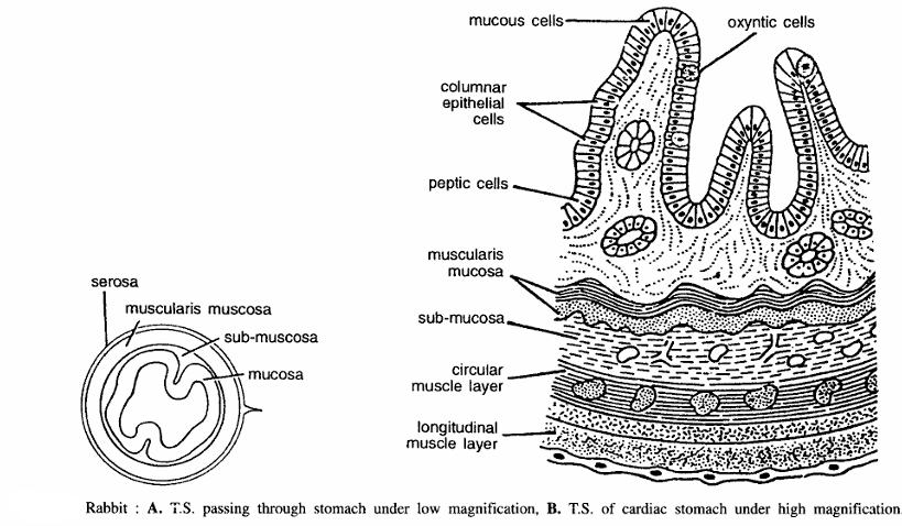
Special features (functions) : By alternate contraction and relaxation of stomach muscles, food is masticated and pushed forwards and digested. Pepsin breaks peptide bonds and converts proteins into derived proteins i.e., peptones and proteoses. Lipase hydrolyses fats into glycerol and fatty acids.
Identification : Since it has thick longitudinal muscles and all above features, hence it is T.S. of cardiac stomach of rabbit.
Mammal Slides Rabbit T.S. Passing Through Ileum
Comments :
- A. Under low magnification: (10 X eye-piece; 4 X objective).
- The T.S. through ileum shows that it is composed of outer serosa, muscular coat, sub-mucosa, muscularis mucosa and mucosa.
- B. Under high magnification : (10 X eye-piece; 40 X objective).
- Serosa fonns outer thin layer covering containing squamous epithelial layer.
- Muscular coat consists of outer longitudinal and inner circular fibres.
- Longitudinal muscle layer is comparatively thinner. By their contraction intestinal tube is shortened but its lumen is widened.
- Circular muscle layer is almost double in thickness than the L.M.L. On its (CML) contraction there is increase in the size of the intestine but decrease in the lumen.
- Sub-mucosa is well developed and is composed of loose connective tissue. Sub-mucosa is very thin layer consisting of outer longitudinal and inner circular muscle layer.
- Mucosa is thrown into villi or folds composed of single-layered endodennal columnar epithelial cells. From the base of villi upto surface layer there are several tubular simple or branched glands called as crypts of Lieberkuhn. These glands are lined by epithelial cells containing goblet cells.
- Above muscularis mucosa there are several nucleated rounded glands called as Brunner’s glands. Villi are composed of tall simple columnar endodennal columnar epithelial cells or absorptive cells and rounded goblet cells. Several nuclei are seen. The inner substance of the villi contains connective tissue, lacteals and nuclei. Along with basal nuclei rounded lymphocyte cells are clearly seen.

Special features (functions) : Ileum receives three kinds of digestive enzymes – (a) pancreatic juice containing trypsin, amylase and lipase, (b) bile juice containing bicarbonate, glycocholate and taurocholate, and (c) succus entericus containing peptidase, lipase, invertase, maltase and lactase. These enzymes are secreted in response to two honnones secreted in duodenum: (i) cholecystokinin which stimulates liver to secrete bile juice, and (ii) secretin which stimulates pancreas to secrete pancreatic juice. In ileum food is converted into amino acids, polypeptides, maltose, glycerols and fatty acids.
Identification : Since villi contain Goblet cells and all above features, hence it is T.S. of ileum (small intestine) of rabbit.
Mammal Slides Rabbit T.S. Passing Through Liver
Comment
- Liver of rabbit is a five-lobed structure. T.S. passing through liver shows hepatic strands, bile ducts, blood vessels and central vein.
- The liver is a solid glandulo-reticular organ made of polyhedral radiating column of cells called as laminae.
- Bile canaliculi lie among the hepatic cells and connect in groups fonning bile ductule or portal-tract consisting of bile, duct hepatic artery and hepatic vein.
- Each hepatic lobule is pierced everywhere with a network of sinusoid.
- Conspicuous cells occur at intervals on the walls of the sinuses. These are called as stellate or Kupffer cells. They are highly phagocytic and they ingest erythrocytes and other suspended particles. Kupffer cells could be best seen under high magnification presence of Kupffer cell indicates immune response.

Special features (functions) : (i) Liver is a very important organ for metabolism and has following functions, (ii) It secretes bile juice consisting of bile salts, bile pigments and lecithin, (iii) It stores glycogen, inorganic salts of iron. copper and basophilic ribo-nucleoprotein, (iv) Glycogenesis and Glycogenolysis take place in liver, (v) Liver produces fibrinogen and prothrombin which are essential components of blood clotting. It also produces heparin, an anticoagulant substance, (vi) Liver changes ammonia into urea by ornithine cycle, (vii) It controls oxidation of sugar, (viii) Various enzymes are synthesized in liver, (ix) It also stores and synthesizes vitamins, (x) In embryonic condition it produces blood corpuscles.
Identification: Since the section shows radiating column of cells and above features, hence it is T.S. of liver of rabbit.
Mammal Slides Rabbit T.S. Passing Through Pancreas
Comments:
- Under low magnification: (10 X eye-piece and 4 X objective).
- Pancreas is a very important digestive gland. T.S. passing through it shows that it is composed of various alveoli or pancreatic acini. It is a compound tubulo-alveolar racemose gland consisting of both exocrine and endocrine parts.
- The mammalian pancreas can be distinguished from that of frog in having distinct lobulations, alveoli or pancreatic acini and islets of Langerhans.
- Each pancreatic lobe contains 10 to 20 secretory cells or acini which are nucleated. The central part has narrow to wide lumen. The pancreatic duct, large artery and vein are also seen in the section. Several cut blood vessels present in connective tissue.
- B. Under high magnification : (10 X eye-piece; 40 X objective).
- Acini and islets of Langerhans are very clearly seen. The wall of each acinus is made up of columnar or pyramidal cells. Each cell contains a central nucleus and course granules. Each acinus has wide lumen. The region of islets of Langerhans reveals 3 or 4 kinds of cells-alpha and beta and undifferentiated cells.

Special features (functions) : Endocrine islets of Langerhans, in eosin-haematoxylin-stained sections, appear as rounded masses of cells with unstained cytoplasm and they contain 3 kinds of cells :
- Alpha cells which secrete a hormone called as glucagon. It increases blood sugar level in the body and its deficiency causes hypoglycemia.
- Beta cells secrete another honnone, insulin, which plays an important role in carbohydrate metabolism. Its deficiency causes diabetes. It regulates blood sugar level,
- Some unknown undifferentiated cells of unknown function. Acini cells secreate 3 kinds of pancreatic enzymes such as : (i) Amylopsin (amylase) which acts on starch and glycogen and changes them into maltose; (ii) Tripsinogen (trypsin) which acts on peptones and proteoses to change them into aminoacids; and (iii) Lipase which hydrolyses fats into fatty acids and glycerols.
Identification: Since the section contains acini and above features, hence it is T.S. of pancreas of rabbit.
Mammal Slides Rabbit : T.S. Passing Through Spleen
Comments :
- Under low magnification: (10 X eye-piece; 4 X objective).
- Spleen is the largest lymphoid gland.
- It is dark brown in colour and composed of muscular and fibrous coat, fibrous trabeculae, red pulp, white pulp and is richly vascularised.
- Capsule sends bands or trabeculae, which form a network in the substance of the gland.
- In the interstices of the framework lies a soft pulpy substance, called as spleen pulp which may be red or white.
- Red pulp forming the bulk gives red colour to spleen.
- Section also contains blood corpuscles, malpighian corpuscles, blood capillaries venous sinuses and nerves.
- Under high magnification : (10 X eye-piece; 40 X objective).
- A portion of the spleen under high magnification shows clear distinction between red and white pulp.
- Other structures seen are penicilli of white pulp and venous sinuses.
- White pulp, contains dense concentration of lymphocytes and looks darker while nuclei of red pulp are scattered. Indistinct reticular and phagocytic cells are also present.

Special features (functions) : Spleen has following functions – (i) Red and white pulp contain resident macrophages, T-Iymphocytes, platelets and B-Iymphocytes. It also contains plasmocytes secreting antibodies, (ii) It stores and synthesizes leucocytes, (iii) It contains macrophages which destroy old erythrocytes. (iv) In embryonic condition it produces erythrocytes but after birth lecucocytes are produced.
Identification : Since the section contains red pulp, white pulp and all above features, hence it is T.S. of spleen of rabbit.
Mammal Slides Rabbit T.S. Passing Through Thyroid Gland
Comments :
- Under low magnification : (10 X eye-piece; 4 X objective).
- It is the most familiar endocrine gland, consisting of right and left lobes connected across to the ventral side of trachea by isthmus.
- Thyroid gland comprises of a framework of connective tissue, enclosing numerous rounded or oval follicles or vesicles of different sizes and covered by capsule.
- Histologically it consists of a Humber of rounded thyroid follicles of various sizes, separated by one another by connective tissue strands.
- Thyroid is richly supplied with blood vessels and nerves. It is innervated from the sympathetic nerves.
- Thyroid secretes thyroxin which contains an amino acid and 65% of iodine.
- B. Under high magnification : (10 X eye-piece; 40 X objective).
- The thyroid is composed of follicular and interfollicular zones. Follicles are surrounded by single layered cuboidal epithelial cells. Lumen of each follicle, contains a viscous liquid called as thyroid colloid. Interfollicular zone contains nerves, blood vessels and large number of nuclei : nerves, blood vessels and large number of nuclei.
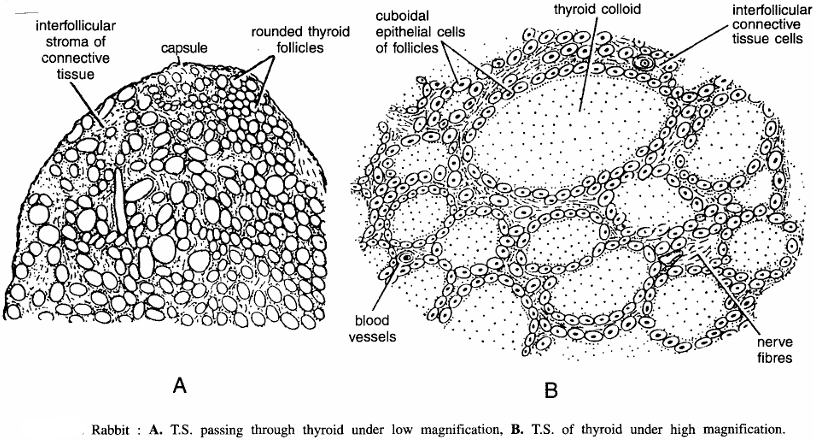
Special features (functions) : Thyroxin controls entire metabolism. Its deficiency causes lowered metabolism. Hyperthyroidism results m protrusion of eye-balls. Removal of thyroid from the frog tadpole stops metamorphosis. Thyroid gland is controlled by TSH (thyroid stimulating hormone) from pituitary.
Identification : Since the section shows rounded follicles and all above features, hence it is T.S. of thyroid gland of rabbit.
Mammal Slides Rabbit : T.S. Passing Through Parathyroid Gland
Comments :
- Two parathyroid glands follicles are always found embedded in the substance of thyroid or found on each side of the thyroid gland.
- Each parathyroid is enclosed within a capsule.
- Each parathyroid is a glandular organ consisting of columns of epithelial cells and blood vessels.
- The gland contains chief cells, colloid or oxyphil cells, separated by the connective tissue.
- Parathyroid produces a hormone called as parathormone, which is devoid of iodine.
- Removal of parathyroid causes tetany or death. Numerous sinusoid blood channels run between the columns.
- Parathyroid gland develops as epithelial outgrowth from the third and fourth branchial clefts of the embryo.

Special features (functions) : The secretion of parathyroid gland controls calcium and phosphate concentrations in the blood plasma and it also controls their metabolism in the body.
Identification : Since the section contains column of cells and all above features, hence it is T.S. of parathyroid gland of rabbit.
Mammal Slides Rabbit : T.S. Passing Through Adrenal Gland
Comments :
- Adrenal glands lie at the anterior end of kidneys and are two in number.
- T.S. passing through the adrenal gland shows that it is composed of outer vertically striated, yellow coloured cortex and inner soft, highly vascular and dark brown medulla.
- The entire gland is covered by fibrous capsule, which sends septa into the cortex and divides it into 4 zones :
- Zona glomerulosa. This zone is found below the capsule. It comprises of columnar cells. This zone controls the mineral and water balance of the body and also fat and carbohydrate absorption. Active hormone is deoxycorticosterone which is not under pituitary control,
- Zona fasciculata. This zone consists of compressed cells which secrete corticosterone to control carbohydrate metabolism. This hormone also causes disintegration of lymphocytes and release of their antibodies,
- Zona reticularis. Next to medulla, this zone consists of pigmented reticular cells. This zone secretes sex hormones.
- Medulla has irregularly disposed cells. It contains elastic fibres and sinusoids (large blood spaces). Medulla cells have granular cytoplasm which take dark stain with osmic vapour.
- In some medulla cells are chromoffin bodies or paraganglia.
- Cortex has mesodermal origin and medulla neuroectodermal, and have abundant blood supply.
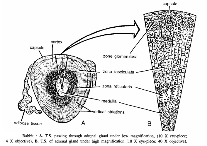
Special features (functions) : Adrenal gland secretes important hormones both from medullary and cortical zones. The medullary hormones are Epinephrine (Adrenaline) and Norepinephrine. Epinephrine is hypoglycemic hormone, promotes glycogenolysis and increases blood pressure. The cortical region secretes corticoids which promote gluconeogenesis and marked effect on protein metabolism.
Identification : Since the section shows 4-zones and all above features, hence it is T.S. adrenal gland of rabbit.
Mammal Slides Rabbit T.S. Passing Through Lung
- Under high magnification: (10 X eye-piece; 40 X objective).
- T.S. through lungs shows bronchioles, alveoli, blood vessels, lymphatic vessels and nerves.
- Bronchi are lined by ciliated epithelium resting with basement membrane.
- Ciliated columnar epithelial cells are surrounded by circular muscle fibres. Next to muscles are loose fibron spongy tissues contammg bronchioles mucous glands and alveoli.
- Alveoli contain 2 winds of (i) small cubical thick cells and (ii) very thin nucleated cells.
- Bronchioles have lumen.
- Several cut pulmonary veins and arteries are observed.
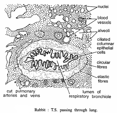
Special features (functions) : Lung is a very important respiratory organ, where atmospheric O2 combines with haemoglobin to form oxyhaemoglobin in blood capillaries. From the lungs oxyhaemoglobin is transported to body for cellular respiration. C02 is also expelled from the body through the lungs.
Identification : Since the section contains alveoli and all above features, hence it is T.S. lung of rabbit.
L.S. Passing Through Kidney
Comments :
- A. Under low magnification: (10 X eye-piece; 4 X objective).
- Kidney is metanephric, compact, bean-shaped, retro-peritoneal, compound, tubular gland, attached to dorsal body wall. Covered by retroperitoneal covering and fibroin capsule.
- Sagittal section of the kidney reveals two distinct portions-(i) cortex, and (ii) medulla. Between these two zones is undefined boundary zone characterized by large blood vessels.
- Cortex has several rounded Bowman’s capsules. Medulla is subdivided into conical portions called pyramids.
- Cortex and medulla are entirely composed of uriniferous tubules, which have straight direction in the medulla and contorted arrangement in the cortex. Renal artery renal vein and ureter enter at the hilum.
- Groups of straight tubules pass front the medulla through the thickness of the cortex forming medullary rays.
- Between medullary rays are the deep cortical down growths, called as renal column of Bertini.
- Under high magnification : (15 X eye-piece; 4 X objective).
- Uriniferous tubules are lined with large granular ciliated epithelial cells and begin in the cortical part of the organ in dilation as Bowman’s capsules, which enclose convoluted tufts of blood capillaries called glomerulus and several nuclei.
- Capsule is lined by flattened epithelium. Glomerulus is formed by branches of afferent and efferent vessels. Tubule leaves the capsule by neck and it forms proximal convoluted ascending limb, descending limb and loop of Henle. Blood vessels are also seen in the section.

Special features (functions). Kidney is the important excretory organ. It filters from the blood water, urea, uric acid and phosphates.
Identification: Since section contains cortex medulla and all above features, hence it is V.S. kidney of rabbit.
T.S. Passing Through Testis
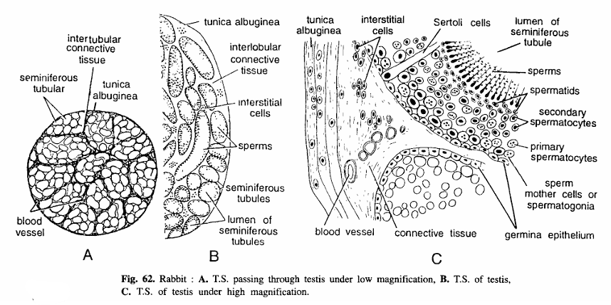
Comments:
- Under low magnification: (10 X eye-piece; 4 X objective).
- There is a pair of smooth, oval-shaped testes, each enclosed In a thin envelope, called as tunica albuginea
- Histologically each testis is internally divided into a number of lobules with occasional internal communications and separated by connective tissue.
- Glandular substance of the testis is wholly made up of convoluted seminiferous tubules. Large number of cut seminiferous tubules are seen with varying diameter.
- Section shows tunica albuginea, cells, sperms, seminiferous tubules and lumen of seminiferous tubules
- Interstitial cells which produce a hormone, called as testosterone. Which is responsible for the development of male secondary sexual character.
- Under high magnification : (10 X eye-piece; 40 X objective).
- Testis is covered by serosa and a fibrous coat or tunica albuginea.
- At the interjection of two seminiferous tubules, connective tissue, interstitial cells vacuoles and blood vessels are seen. In the seminiferous tubules are some nutritive Sertoli cells. Seminiferous tubules appear rounded or oval in section. Each tubule is surrounded by a thin basement membrane lined by germinal epithelium.
- From basement membrane to inwards there are several kinds of cells: (i) Spermatogonica lie along periphery of tubule and appear closely packed together, (ii) Primary spermatocytes – They have the largest cells and large nuclei, (iii) Secondary spermatocytes – Smaller cells with deeply stained nuclei, (iv) Spermatids – Small clusters of cells with condensed nuclei, (v) Spermatozoa or sperms lie in the cavity of the tubule.
- Sperm has head and tail.
- The nucleus of the sperm lies in the head which is pointed as the acrosome.
- Outer covering tunica albuginea interstitial cells and blood vessels etc. are seen in the section T.S. testis of rabbit.
Identification: Since the section contains sperms and all above features, hence it is T.S. testis of rabbit.
Rabbit : T.S. Passing Through Ovary

Comments:
- Under low magnification: (10 X eye-piece; 4 X objective)
- Outer most layer is of peritoneum which has cupical cells.
- Just beneath peritoneum is germinal epithelium bounded by connective tissue called as tunica albuginea.
- Germinal epithelium gives rise to oogonia, developing follicles and Graafian follicle.
- Section shows young follicles and mature Graafian follicles and corpus luteum.
- Interior of the section shows connective tissues, interstitial cells and blood vessels.
- Under high magnification : (10 X eye-piece; 4 X objective).
- Detailed structure of Graafian follicle in seen under high magnification. Follicle is surrounded by connective tissue or stroma.
- Fully mature oocyte is surrounded by a thick transparent layer called Zona pellucida surrounded by another layer corona radiate.
- Corona radiata in surrounded by mass of cells called as discus proligerous or cumulus.
- Corona radiale is surrounded by liquor folliculi and then by membrane granulosa. Thick membrane granulosa in covered by thick layer called as theca folliculi.
Identification : Since the section contains Graafian follicle and all above features, hence it is T.S. of ovary of rabbit.
T.S. Passing Through Bone

Comments :
- Bone in T.S. in covered by thin layer called as periosteum.
- Below periosteum is endosteum.
- Space in endosteum is filled by matrix which contains periosteal lamellae and interstitial lamellae.
- Interstitial lamellae contains Haversian system. Haversian system is composed of central Haversian canal surrounded by concentric rings of lacunae and canaliculi.
- Each Haversian contains blood vessels, lymph vessels and nerves.
Identification : Since the above section contains Haversian canal and above features, hence it is T.S. of bone of rabbit.
T.S. Passing Through Placenta
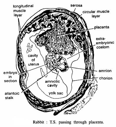
Comments :
- Structure of placenta shows outer serosa, longitudinal and circular muscle fibres, uterine crypts.
- In placenta foetal villi and uterine crypts are seen.
- In section embryonic part in broad.
- Chorion, amnion, amniotic cavity, yolk sac, allantoic stalk, embryo in section and cavity of uterus seen.
- It is discoidal placenta of haemochorial type. In blood vessels of chorionic epithelium and foetal villi maternai blood directly flows.
Identification : Since the section contains embryo and all above features, hence it is of placenta of rabbit.
T.S. Passing Through Spinal Cord
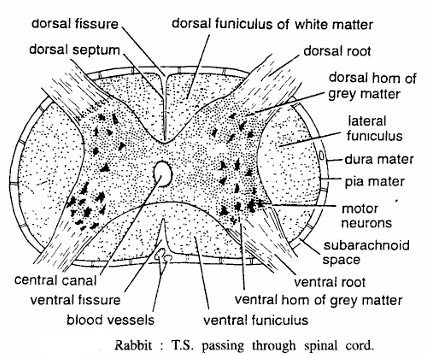
Comments :
- Just after emergence from the foramen magnum, medulla continues as spinal cord.
- T. S. of spinal cord shows that it is covered by three membranes outer piameter, middle arachnoide space and inner durameter.
- Entire nervous tissue is divided into outer white matter and inner grey matter.
- White matter and grey matter are made up of nerve cells and nerve fibres respectively.
- Grey matter is H-shaped or butterfly shaped and is perforated by a central canal.
- Grey matter projects dorsolaterally and ventrolaterally as paired dorsal and ventral horns respectively for attachment of nerve roots.
- Dorsal and ventral horns penetrate deep into white matter as dorsal and ventral septums respectively. Other structures seen are motor neurons and blood vessels.
- Spinal cord gives spinal nerves which control reflex activities and also conduct impulses to and pro from brain,
Identification : Since the grey matter is H-shaped a butterfly shaped and shows above features, hence it T.S. of spinal cord of rabbit.
Image References :- Practical zoology Vertebrate