
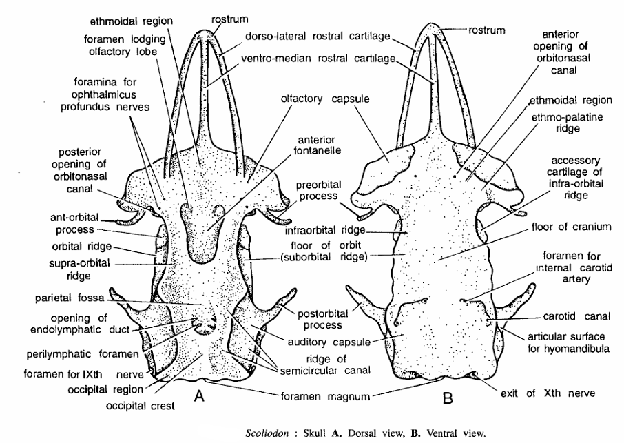
Skull Bones of Scoliodon
Skull Bones are consists of
- Cranium and sense capsules
- Visceral skeleton.
Comments
1. Cranium and sense capsules
Made up of a single piece of cartilage and divisible into 4 regions.
- Occipital region : Posterior-most part having a median opening, the foramen magnum. Lateral sides contain occipital condyles and occipital crest in the middle.
- Auditory region : Lies in front of occipital, in the form of two auditory capsules one on each side. Each capsule encloses internal ear. Protuberances for semicircular canals can be seen. This region dorsally contains openings for endolymphatic duct and perilymphatic foramen. Below postorbital process, articular surface for hymandibula of hyoid arch is seen.
- Orbital region : Orbital region contains eye during life time. Each orbital region is made up dorsally by supra-orbital ridge, ventrally by infra-orbital ridge, anteriorly by pre-orbital process and posteriorly by post-orbital process. Number of foramina or nerves are found within orbit.
- Ethmoidal region : It includes the anterior part of cranium, two nasal or olfactory capsules and rostrum. Rostrum is made by a ventro-median rostral-cartilage and two dorsolateral rostral cartilages.
- Other structures seen in dorsal view are foramen lodging olfactory lobe, foramen for ophthalmicus profundus nerves, posterior opening of orbitonasal canal, anterior orbital process, parietal fossa, foramen for IXth nerve. Occipital crest, ridge of semicircular canal and anterior fontenelle.
- Structures seen in ventral view are rostrum, anterior opening of orbitonasal canal ethmopalative ridge, accessory cartilage of infra-orbital ridge, floor of cranium, foramen for internal carotid artery, carotid canal, and exit of IXth nerve.
2. Visceral skeleton
It forms jaws, skeleton of pharynx and a series of V-shaped visceral arches around buccal cavity and pharynx. Important arches are :
- First or mandibular arch. It is made up of two segments. Dorsal segment is called as palato pterygoquadrate forming upper jaw. Lower segment is called as Meckel’s cartilage forming lower jaw.
- Second or hyoid arch. It consists of basihyal, ceratohyal and hyomandibular. Jaws are suspended through hyomandibula and this is called as hyostylic suspensorium.
- Branchial arches. Remaining 5 visceral arches are known as branchial arches. Each branchial arch consists of 4 rod-like pieces : dorsal pharyngobranchial, lateral epibranchial and ceratobranchial and ventral hypobranchial. Only epibranchials and ceratobranchials bear gill rays supporting gills.
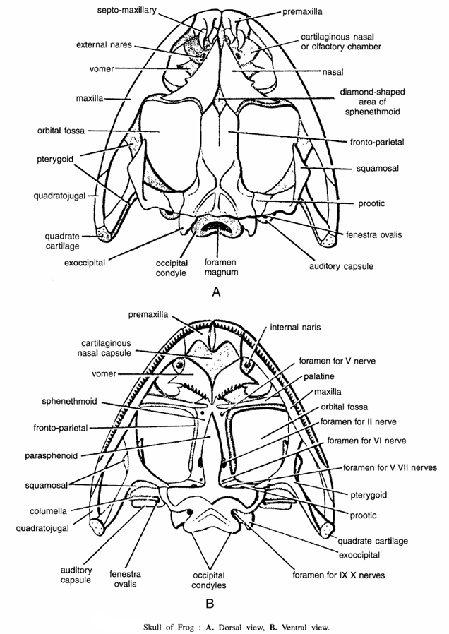
Skull Bones of Frog
- Dorsal view : Various structures seen are septo-maxillary, external nares, vomer maxilla, orbital fossa, pterygoid, quadratojugal, quadrate cartilage, exoccipital, occipital condyle, foramen magnum, auditory capsule, fenestra ovalis, prootic, squamosal, fronto-parietal, diamond-shaped area sphenethmoid, nasal, cartilaginous nasal or olfactory chamber and premaxilli.
- Ventral view : Various structures seen are premaxilla, cartilaginous nasal capsule, vomer, sphenethmoid, fronto parietal, parasphenoid, squamosal, columella, quadratojugal, auditory capsule, fenestra ovalis, occipital condyle, foramen for IX and X nerves, exoccipital quadrate cartilage, prootic, pterygoid, foramen for V and VII nerves, foramen for VI nerve, foramen for II nerve, orbital fossa, maxilla, palatine, foramen for V nerve and internal nares.
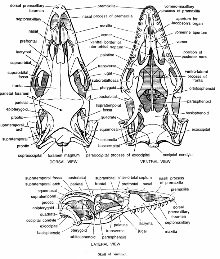
Skull Bones of Varanus
- Dorsal view : Various skull bones seen are as follows- Dorsal premaxillary foramen, septomaxillary nasal, prefrontal, lacrymal, supraorbital fossa, frontal parietal foramen, parietal, epipterygoid, prootic, supratemporal arch, supratemporal, prootic, supraoccipital, foramen magnum, columella, squamosal, quadrate, supratemporal fossa, postorbital, pterygoid jugal, transverse, palatine, ventral bogeler of inter orbital septum, vomer, maxilla, nasal process of premaxilla, premaxilla and paraoccipital process of exoccipital
- Ventral view : Various skull bones in ventral view consists of premaxilla, maxilla, vomer, ventral borae;’ of inter orbital septum, palatine, transverse, jugal, sub-orbital fossa, pterygoid, postorbital, quadrate, squamosal, columella, basioccipital, paraoccipital process of exoccipital, occipital condyle, ex occipital, basisphenoid, parasphenoid, orbitosphenoid, ventrolateral process of frontal, position of posterior nare, vomer, vomerine aperture, apparatus for Jacobson’s organ and vomeromaxillary process of premaxilla.
- Lateral view : Various skull bones seen in lateral view consists of premaxilla, nasal process of premaxilla, nasal, prefrontal, interorbital septum, frontal, supraorbital, parietal, postorbital, supratemporal fossa, supratemporal arch, squamosal,’ supratemporal, prootic, epipterygoid, quadrate, occipital condyle, exoccipital, basisphenoid, pterygoid, orbitosphenoid, parasphenoid, transverse, palatine, jugal, lacrymal, maxilla, septomaxillary, dorsal premaxillary foramen, premaxilla.
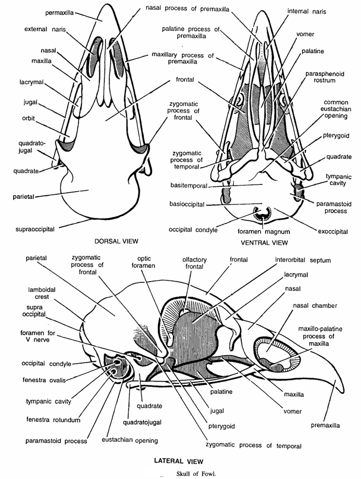
Skull Bones of Fowl
- Dorsal view : Various skull bones seen are as under-premaxilla, external nares, nasal, maxilla lacrymal, jugal, orbit, quadratojugal, quadrate, parietal, supraoccipital, zygomatic process of frontal, frontal, maxillary process of premaxilla, palatine process of premaxilla and nasal process of premaxilla.
- Ventral view : Various skull bones seen are nasal process of premaxilla, palatine process of premaxilla, maxillary process of premaxilla, frontal zygomatic process of frontal, zygomatic process of temporal basitemporal, basioccipital, occipital condyle, exoccipital, paramastoid process, tympanic cavities, pterygoid, common eustachian opening, parasphenoid rostrum, palatine vomer and internal naris.
- Lateral view : Various skull bones seen in lateral view consists of maxillopalatine process of maxilla, nasal chamber nasal lacrymal, inter-orbital septum, frontal, olfactory frontal, optic foramen, zygomatic process of frontal parietal lamboidal crest, supraoccipital foramen of V nerve, occipital condyle, fenestra ovalis, tympanic cavity, fenestra rotundum, paramastoid process, eustachian opening, quadratojugal, quadrate, zygomatic process of temporal, pterygoid, jugal, palatine, maxilla, vomer and premaxilla.
Skull Bones of Rabbit
- Dorsal view : Skull bones seen in dorsal view are anterior nares, pre-maxilla, nasal, maxilla, frontal, jugal, parietal, squamosal, tympanic bulla, interparietal, supraoccipital, external auditory meatus, zygomatic process of squamosal, supra-orbital process of frontal, zygomatic arch, zygomatic process of maxilla, maxillary process of frontal, nasal or frontal process of premaxilla and I incisor tooth.
- Ventral view : Skull bones seen in ventral view are incisor tooth, palatine process of pre-maxilla, anterior palatine foramen, palatine process of maxilla, premolar teeth, zygomatic process of frontal, zygomatic process of squamosal, eustachian canal, external auditory meatus, para-occipital process of exoccipital, supraoccipital, foramen magnum periotic, occipital condyle, exoccipital, mastoid process, tympanic bulla, basioccipital, alisphenoid, basisphenoid, pterygoid, pituitary foramen, presphenoid, palatine, vomer, maxilla, pre-maxilla, nasal and II incisor tooth.
- Lateral view : Skull bones seen in lateral view consists of premaxilla, I-incisor, II-incisor, zygomatic process of maxilla, premolars, molars, jugal, palatine, pterygoid, basisphenoid, basioccipital, tympanic bulla, paraoccipital process of exoccipital, occipital condyle, stylomastoid foramen for facial nerve, external auditory meatus, periotic, supraoccipital, interparietal, post tympanic process of squamosal, squamosal, zygomatic process of squamosal, parietal, alisphenoid, sphenoid fissure, supraoccipital process of frontal, frontal, optic foramen, orbitosphenoid, lacrymal, maxillary process of frontal, nasal or frontal process of premaxilla.
