Comparative study of Hind limb bones of vertebrate
The hind limb of vertebrates have a homologous five-toed plan – femur, tibia/fibula, tarsus, metatarsals and phalanges – adapted to specific modes of locomotion. Structural variations exist: running mammals (horses) have reduced toes (cannon bones) to increase speed, birds have a fused tibia, while tetrapods like salamanders retain primitive structures. The hind legs are generally stronger than the front legs.
Hind Limb Bones of Frog
Hind limb constitute the femur, tibio-fibula, astragalus-calcaneum and bones of foot.

Comments
- Femur
- It is thigh bone having expanded ends, covered with calcified cartilages.
- Proximal end or head articulates with acetabulum while distal end with tibio-fibula.
- Tibio-fibula
- These are shank bones and are elongated.
- Tibia is inner and fibula is outer, both fused together to form a compound bone.
- Proximally tibia contains a tibial crest.
- At proximal end tibio-fibula articulates with femur, and at distal end with astragalus-calcaneum. Structures seen are proximal articular facet, groove, tibia, fibula, nutrient foramen, distal groove and distal articular facet.
- Astragalus-calcareum
- These are proximal ankle bones united at both ends and covered by proximal and distal epiphyses of calcified cartilages at proximal and distal ends respectively.
- Outer thicker straight bone is calcaneum of iibulare, while inner thinner and slightly curved bone is astragalus or tibiale.
- Bones of foot
- Foot contains 5 long, slender bones, known as metatarsals, having five true toes.
- First, second, third, fourth and fifth digits contain 2, 2, 3, 4 and 3 phalanges respectively.
- Sixth toe is very small having 2 or 3 bones and is called as pre-hallux or calcar.
Hind Limb Bones of Varanus
Hind limb consists of femur, tibia, fibula and bones of hind foot.
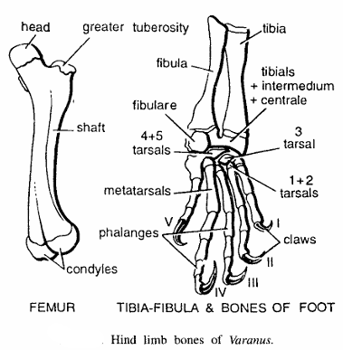
Comments:
- Femur
- It is thigh bone having two epiphyses.
- Proximal end contains head, which fits into acetabulum, while distal end is pulley-shaped, having two tuberosities or for articulation with tibia and fibula,.
- Femur condyles has lesser trochanter and greater trochanter on pre-axial and post-axial sides, respectively.
- Tibia and fibula and bones of hind foot
- These are shank bones
- Tibia is stout, curved and on pre-axial side, while fibula is slender and on post-axial side.
- Bones of hind-foot
- It is made up of 5 tarsal bones.
- First, second, third, fourth and fifth toes contain 2, 3, 4, 5 and 3 phalanges, respectively.
- Each toe bears a terminal horny claw.
Hind Limb Bones of Fowl
Hind limb comprises of femur, tibio-tarsus and fibula, tarsals, tarso-metatarsus and phalanges. Hind is modified for bipedal locomotion.
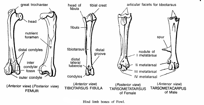
Comments:
- Femur (Anterior view)
- It is cylindrical, short and curved thigh bone.
- Head fits into acetabulum and outwardly it contains greater trochanter.
- Distal end is pulley-shaped for patella bone and bounded by two condyles to articulate with tibia namely outer condyles and inner condyle with inter condylar fossa.
- Femur (Posterior view)
- Contains nutrient foramen, head, inter condylar fossa, outer condyle and inner condyle.
- Tibio-tarsus and fibula (Anterior view)
- Tibio-tarsus and fibula form shank bones.
- Tibio-tarsus is a straight and long bone formed by fusion of tibia with proximal row of tarsals (astragalus and calcaneum).
- Proximally, it articulates with femur and distally with tarso-metatarsus.
- Fibula is reduced to a small slender bone. Other structures are tibial crest distal lateral tubercle, distal groove.
- Tarso-metatarsus female (Posterior view).
- It is a compound bone formed by fusion of distal row of tarsals with second, third and fourth metatarsals. Other structures seen at proximal end are articular facets for tibiotarsus, nodule of I metatarsal, II metatarsal, III metatarsal and IV metatarsal.
- Tarso-metatarsus. Male (Anterior view). In males tarsometatarsus contains a spur. Phalanges.
- Four toes.
- First or hallux is directed backwards and remaining three forwards.
- First, second, third and fourth toes have 2, 3, 4 and 5 phalanges respectively; each one is clawed.
Hind Limb Bones of Rabbit
Hind limb is formed by femur, tibio-fibula and bones of hindfoot.

Comments :
- Right femur (Back view)
- It is thigh bone.
- Proximal head articulates with acetabulum.
- Lesser, greater and third trochanters present for muscle attachment.
- Distally it has pulley-shaped structure, having two lateral condyles which enclose an intercondylar groove.
- Right femur (Front view) :- It shows head, greater trochanter, lesser trochanter, shaft, patellar groove and condyles.
- Bones of hind foot :
- It contains tarsal bones in two rows.
- Tibiale and intermedium of the proximal row are fused to form astragalus on pre-axial side, while fibulare or calcaneum is the largest tarsal bone produced into a spur on post-axial side.
- Distal row contains three bones mesocuneiform, ectocuneiform and cuboid.
- Only four toes each having three phalanges, the terminal one bearing a claw.